Is it Just a Bowl Of Cereal?

Ethics in Medical Practices: Part 1

By Krista Surprenant


Quaker Oats Advertisement – 1901

Most of us have experienced a health kick at some point in our lives—reading nutrition information, counting calories, and tracking vitamin levels to hit the perfect daily intake. Maybe you’ve even looked up the health benefits of your favorite products. But have you ever stopped and asked yourself how those nutrient levels were determined?

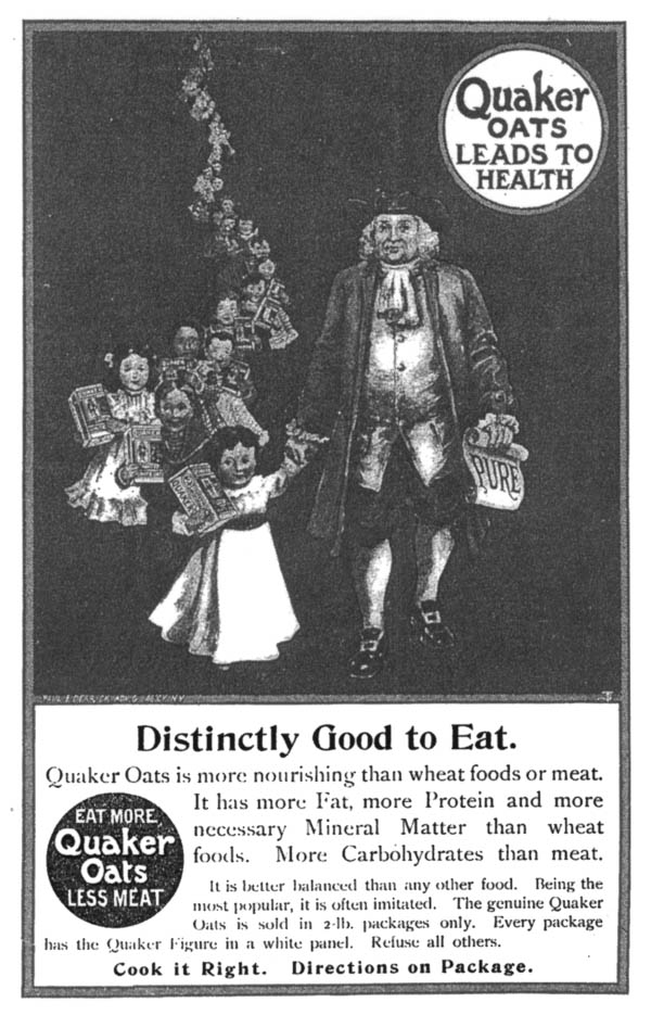
For one brand, information on nutrient levels can be traced back ~75 years to a specific study done at The Massachusetts Institute of Technology (MIT)1. Prior to MIT’s experiments in 1949, studies showed an acid found in oats had a negative impact on calcium, iron, and phosphorus absorption, sparking debate around the nutritional value of oat-based cereals [1,5]. This raised consumers’ concerns about major brands such as Quaker Oats. In order to defend their product against competitors like Cream of Wheat, Quaker Oats funded MIT’s study to track iron and calcium absorption following cereal consumption [1]. The company hoped to demonstrate that their cereal did not reduce mineral absorption and was nutritionally comparable to Cream of Wheat [1,5].

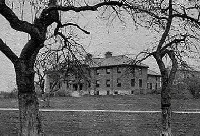
To carry out this experiment, MIT needed volunteers, so they turned to The Fernald State School in Waltham Massachusetts [1,5].

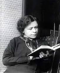
In 1848, the Massachusetts School for the Feeble-Minded was founded by Samuel Gridley Howe with the goal of educating disabled adolescents to become high-functioning, independent individuals [6]. Students were taught academic, vocational, and personal skills, including sewing, housekeeping, music interpretation, and athletics [6]. Over time, the school expanded to house adults requiring custodial care and individuals with more severe disabilities. In 1888, Walter E. Fernald became superintendent of the school and shifted the institution’s philosophy towards a scientific framework rooted in eugenics [6]. Eugenics sought to promote “desirable” traits through selective breeding while eliminating “undesirable” traits—possessed by groups that included people of color, immigrants, and individuals with developmental or intellectual disabilities [6,7]. Those deemed “undesirable” were often institutionalized, including residents of what became known as the Fernald State School in 1925 [6].

During this same period, IQ testing became widespread, and children scoring below average were labeled “morons” or “intellectual delinquents” and frequently institutionalized without parental objection [6,7].

Fernald State School Dormitory for Boys

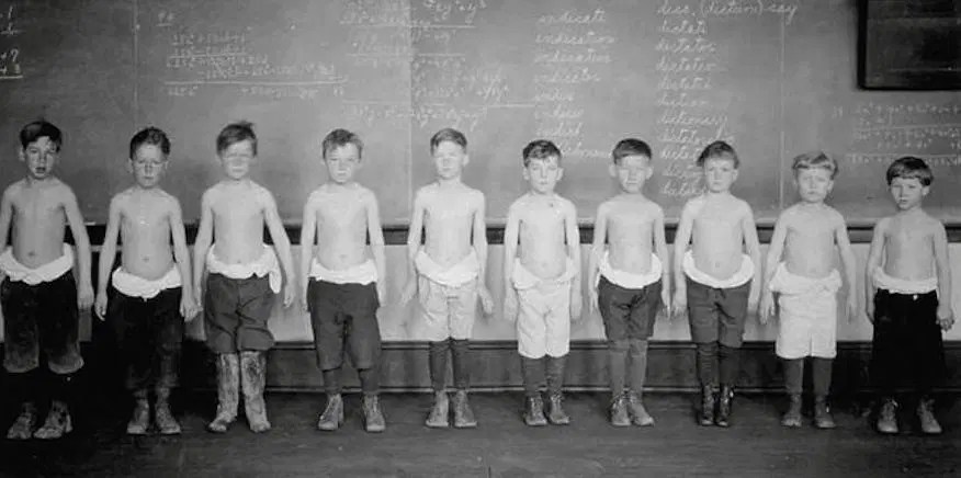
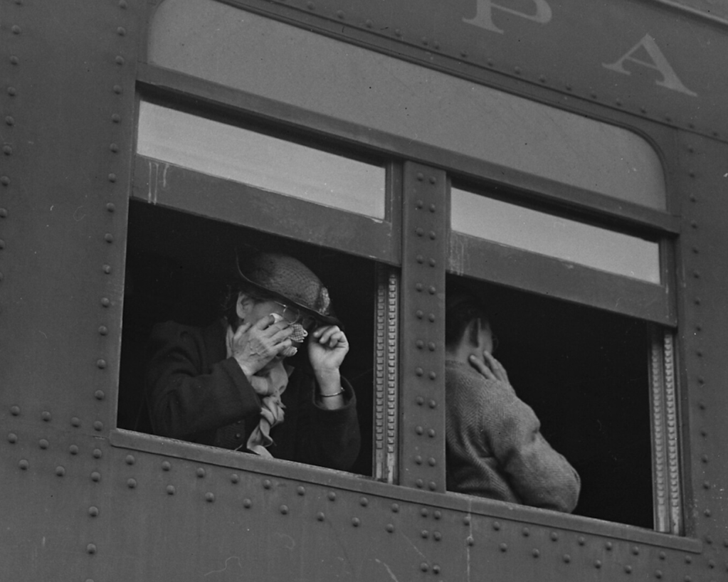
Due to the increased population of students admitted to the Fernald State School, the institution became overrun and underfunded [5,6]. Eventually, the school became home to 2,000-2,500 disabled and unwanted children who were, at the time, considered “not part of the human species” [5,7]. When it opened in 1848, the school’s aim was to provide students with a stable education and sustainable life skills so they could eventually be able to provide for themselves, and be morally rehabilitated (due to thinking disabilities are outward manifestations of corrupt souls). Over time, however, this educational facility that had originally provided nurturing care and solid foundations became a neglectful, abusive custodial institution. Eventually, due to inadequate funding, the incarcerated students who were not physically disabled were used as sources of manual labor to keep the institution functioning without hiring more employees. Years later, at a news conference, one past student at the institution stated, “Instead of being taught, we were being used, and I don’t think that was right. I don’t have the education that a lot of students have now because of my upbringing here. I wasn’t taught anything, I have trouble reading, I can’t spell, I try to get help, and I can’t get help” [5].

This created a perfect opportunity for exploitation of the students.

Because of the large numbers of easily obtainable subjects, MIT recruited disabled students from the Fernald School for an exclusive “science club” at the university. The students were enticed to participate through bribery. Science Club members were offered special privileges including beach trips, better dinners, and access to sporting events. In return for these privileges, the students would be required to participate in various scientific studies, such as the Quaker Oats nutrient tracer study [1,5].

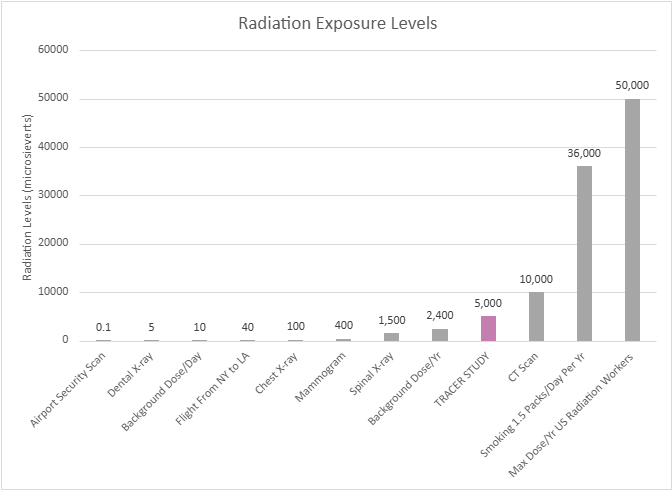
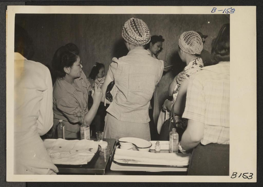
In total, the MIT tracer study used 40 students, ages 7 to 17, to conduct three tracer experiments with the goal of understanding how the body absorbs minerals. The first experiment involved a few dozen students who were fed Quaker Oats cereal coated with small amounts of radioactive iron [1,12]. The radioactive iron chemically behaves just as natural iron would in the body, but its radioactive nature allows it to be traced. In order to trace the iron, the scientists collected frequent blood, urine, and/or stool sample from the participants, measuring the radioactive emissions using a special detector [1,7]. This study revealed that the absorption rate of iron from rolled oats was no less than farina (Cream of Wheat) [1]. The second experiment involved 36 students who received two breakfasts each [1,2]. The milk given to them was spiked with a radioactive calcium tracer, similar to that of the iron, to understand if the phthalate compounds in the oats interfered with dietary uptake of calcium from the milk. The calcium tracer study showed that absorption of a sufficient amount of calcium would not be significantly affected by the phthalates in the oats. The final experiment involved nine students who had radioactive calcium injected directly into their bloodstream to serve as a baseline for how calcium is absorbed in the body without any interference from food or digestion [2].

“Instead of being taught, we were being used, and I don’t think that was right”

The research done by MIT for this study was very well controlled, researched, and executed from a scientific rigor standpoint [1,2]. Therefore, the studies are still considered major scientific discoveries. However, serious ethical issues underlie these ground-breaking studies [5,7]. Due to the state of the Fernald School, operating as a corrupt, underfunded, and apathetic institution, the boys living there were easy targets, vulnerable to coercion with food and fun and ill-equipped to question the studies that would be done at MIT. The parents were not informed either, and were not in a position to intervene. Most of the boys participating in the study were wards of the state2 , therefore the state, not the parents, were their legal guardians. In addition to this, informed consent laws to protect human research subjects did not exist at this time, especially for disabled individuals [7,10]. Consequently, informed consent was not provided to the participants or any living parents. The superintendent of the Fernald School was able to make medical decisions on behalf of the students [5].

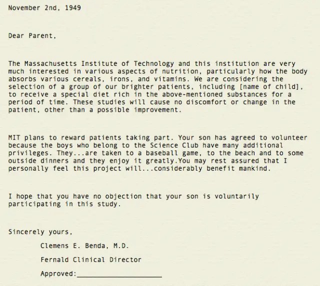
Nevertheless, the school did provide parents with two very deceptive letters, which briefly explained what their child would be participating in, but did not ask for approval. These letters did not give any details of the study, did not mention experiments with radioactivity, nor did they state the possible risks associated with their child’s participation [11]. These letters further alluded that consent from the parent was accepted unless otherwise stated, in an effort to protect the school from any accusations of wrong-doing. Although unethical,these experiments were perfectly legal at that time, due to the lack of modern-day ethical and medical guidelines 3 [7,10]. Interestingly, the researchers conducting this study claimed that disabled children consumed larger amounts of cereal than the average juvenile, to justify their choice to use the boys from Fernald [1,11].

First letter written to inform parents of MIT tracer study – 1949

“Some of us had to sign our own forms, but at that particular time I could not read or write. I had no knowledge of anything other than the fact that I do what I’m told when I’m told.”

– Austin LaRoque, Fernald State School Student

In 1993, almost 40 years after the conclusion of the tracer studies, MIT employed a task force to follow up on the research participants and determine if there were any lasting effects [3]. During the time of participation, the boys involved in the studies experienced levels of radiation equivalent to 50 chest x-rays [3,8]. Luckily, no severe effects were found. It is now known that the amount of radiation the boys experienced was less than current legal regulations. While participants didn’t experience any health complications, their safety had been jeopardized without their knowledge or their parents’. Therefore, the MIT task force suggested, “…all participants who were involved in human subject research which used radioactive materials at Massachusetts-operated facilities for persons with mental retardation should be compensated for any and all damage incurred as a result of such research” [4,9].

Radiation exposure levels in comparison to Tracer Study radiation exposure

MIT chose not to act upon the task force’s suggestion until 1997, when a class action lawsuit for $60 million was filed against MIT and Quaker Oats by study participants and their families, arguing it was a violation of their civil rights. Both MIT and Quaker Oats agreed on a settlement of $1.85 million to each participant to “avoid the expense and diversion of a lengthy legal battle” [4,9]. In addition, MIT’s research director, Dr. Bertran Brill stated, “I admit they shouldn’t have focused on a population that was so captive and had no alternative. That was wrong” [9]. With this statement, the students at Fernald State School were finally indemnified for what had been done to them as children.

Unfortunately, this is not the only time vulnerable groups were targeted for scientific experimentation. From disabled boys, to prisoners, to minority and economically troubled groups, targeting powerless individuals is a shameful trend in the history of science. Even if the studies themselves are scientifically sound, acknowledging the unethical decisions surrounding these studies is very important . Respecting the human rights of all individuals is far more important than any scientific discovery. It is essential that we learn from our past, so that the next generation of scientists will never allow exploitation such as this to happen again.


  1. DISCLAIMER | This study in no way reflects the current ethical decisions of Massachusetts Institute of Technology, Quaker Oats, or any associated studies. ↩︎
  2. A juvenile or incapacitated adult placed under the care of the government due to orphancy, abandonment, or inability to care for the child or for oneself ↩︎
  3. 1947 – Nuremberg Code introduced the ides of voluntary informed consent (not U.S. law, but influenced enactment)
    1964/1975 – Declaration of Helsinki created to serve as the international ethics code for medical research, requiring informed consent, independent review, and protection of vulnerable groups (not binding U.S. law but heavily shaped it)
    1974 – National Research Act was passed in the U.S. and served to require Institutional Review Boards (IRBs) ethics committees and protect people in federally funded research
    1979 – Belmont Report passed as a foundational ethics document explaining respect for persons/informed consent, beneficence to minimize harm, and justice to ensure fair subject selection, explaining why the laws should exist
    1991/2018 Revision – Common Rule for Federal Policy for Human Subjects Protection is the core rule that requires informed consent with full explanation of risk, IRB approval, and gives further protection to children, prisoners, pregnant persons, and people with impaired-decision making, allowing for the monitoring of ongoing studies and ability to withdraw at any time ↩︎

REFERENCES

1 | Boissoneault, L. (2017, March 8). A Spoonful of Sugar Helps the Radioactive Oatmeal Go Down. Smithsonian Magazine. https://www.smithsonianmag.com/history/spoonful-sugar-helps-radioactive-oatmeal-go-down-180962424/

2 | Bronner, F., Harris, R. S., Maletskos, C.J., & Benda, C. E. (1956). Studies in Calcium Metabolism; the Fate of Intravenously Injected Radiocalcium in Human Beings. The Journal of Clinical Investigation35(1), 78–88. https://doi.org/10.1172/JCI103254

3 | Campbell, K. D. (1994, May 11). Task Force Finds Fernald Research had no Significant Health Effects. MIT News | Massachusetts Institute of Technology. https://news.mit.edu/1994/fernald-0511

4 | Court Approves Fernald Settlement. MIT News | Massachusetts Institute of Technology. (1998, January 7). https://news.mit.edu/1998/fernald-0107

5 | Crockett, Z. (2022, March 20). The Dark Secret of the MIT Science Club for Children. Priceonomics. https://priceonomics.com/the-mit-science-club-for-disabled-children/

6 | Daly, M. E. (n.d.). History of the Walter E. Fernald Development Center. https://www.city.waltham.ma.us/sites/g/files/vyhlif12301/f/file/file/fernald_center_history.pdf

7 | Denny, S. (n.d.). Advisory Committee on Human Radiation Experiments Final Report. US Department Of Energy. https://ehss.energy.gov/ohre/roadmap/achre/chap7_5.html

8 | McCandless, D., & Hancock, M. (2013, August). Radiation Dosage Chart. Information is Beautiful. https://informationisbeautiful.net/visualizations/radiation-dosage-chart/

9 | MIT Announces Settlement of Fernald Nutrition Studies Suit. MIT News | Massachusetts Institute of Technology. (1997, December 30). https://news.mit.edu/1997/fernald

10 | Office for Human Research Protections (OHRP). (2026, January 15). Federal policy for the Protection of Human Subjects ‘Common Rule. HHS.gov. https://www.hhs.gov/ohrp/regulations-and-policy/regulations/common-rule/index.html?utm_source=chatgpt.com

11 | Sharav, V. (2016, April 20). MIT reviews Fernald nutrition studies; Vest expresses concern. MIT News | Massachusetts Institute of Technology. https://ahrp.org/1944-1956-radioactive-nutrition-experiments-conducted-by-harvard-and-mit-on-disabled-children/?utm_source=chatgpt.com

12 | West, D. (1998, January). Radiation experiments on children at the Fernald and wrentham schools: Lessons for protocols in human subject research. Accountability in research. https://pubmed.ncbi.nlm.nih.gov/11660586/


IMAGE REFERENCES

1 | Digital Collections – University at Buffalo Libraries. (1901). Advertisement for Quaker oats [Photograph]. Public Domain. Retrieved from https://digital.lib.buffalo.edu/items/show/95016

2 | Fernald State School and Hospital. (Date Unknown). North Building — Historic images and photo gallery. Public Domain. Retrieved from https://fernaldstateschool.com/buildings/north

3 | Human Eugenics Records Archive. (1922). Photograph of Fernald boys in nutrition experiments. Public Domain. Retrieved from http://www.eugenicsarchive.org/html/eugenics/static/images/864.html

4 | Crockett, Z. (1949, 2016). The Dark Secret of the MIT Science Club for Disabled Children [Letter reproduced]. Priceonomics. Public Domain. Retrieved from https://priceonomics.com/the-mit-science-club-for-disabled-children/

5 | Self-Produced. (2026). Information Retrieved from https://informationisbeautiful.net/visualizations/radiation-dosage-chart/

Dr. Kazue Togasaki: Serving the Japanese Community From San Francisco to the Concentration Camp

Manzanar sentry tower Photo by Sonora Ortiz. National Register of Historic Places: 76000484. Public domain.

Prologue

The wail of a newborn broke the silence in the plain, crowded room. Dr. Kazue Togasaki finally smiled in relief and wiped sweat off her forehead. This was her second delivery of the day. Fatigue overwhelmed her, but the parents’ shining faces lifted her remaining strength.

As Dr. Togasaki stepped outside, it was not into the busy streets of San Francisco that she was used to. Instead, a desert wind pushed forcefully into her face. Watchtowers with armed guards stood beyond a few logging camps surrounded by barbed wire.

The towers cast their shadows over the camps, reminding the people there that this was not their home, but Manzanar, one of many Japanese concentration camps built on the soil of the United States.

This was a place where freedom was fenced, and human rights were transgressed.

I had the head nurse and the instructor of nurses at Stanford University Hospital and a doctor, all Japanese, working with me at Tanforan [A temporary detention center located in San Francisco for relocating internees to further remote areas.] — Dr. Togasaki [3].

Childhood and Early Years

You might wonder how Dr. Kazue Togasaki, an American citizen and physician, could end up detained and working in a concentration camp. To answer this, we must begin decades earlier. Dr. Kazue Togasaki was born in San Francisco in 1897 to Japanese immigrant parents [1]. Her father, Kikumatsu Togasaki, who first studied law in Japan, came to the US intending to pursue law in America.

However, social barriers prohibited Kikumatsu from practicing law in the United States. Fortunately, after staying in the US for a while, he met a wonderful woman named Shige Kushida, and they later married in Japan. Then, the couple moved back to the US and chose to pursue a small business.

Even though Kikumatsu Togasaki still thought of himself as a lawyer at heart, Shige, raised in a merchant family, taught him how to run a shop. Dr. Kazue Togasaki recalled, “My parents had a little store at 405 Geary that sold Japanese tea and rice and chinaware…Mother was the daughter of a merchant, so she knew how to buy and sell, but my father, a lawyer, not a businessman…” [3]. Over the years, their family grew to nine children; Dr. Togsaki was the second child and the eldest daughter [3].

Despite being born in the US, Dr. Togasaki did not grow up with the same schooling as many “Caucasian” children. In 1906, the city of San Francisco confirmed the “Exclusion Act” that segregated Japanese children to the “Oriental School.”

Dr. Togasaki was moved from school to school more than once [4]. She said, “The first I knew of discrimination was in 1908 when I was caught in the Exclusion Act” [10]. In the same year of 1906, when Kazue Togasaki was nine, catastrophe struck: One of the most devastating earthquakes in California history killed more than 3,000 people, torching the whole city of San Francisco [5].

Fire in San Francisco following the great earthquake of 1906. View from Gold Gate Park, Marin County, California. (USGS). 1906. Author George R. Lawrence. Public Domain.

I remember we got dressed and walked from our house to the 14th and Church, where there was a hill, and that afternoon and for two days in the daytime we sat, watching the city burn. — Dr. Togasaki [3].

The 1906 California Earthquake changed Dr. Togasaki’s life as a young girl. She saw the terror of the disaster, but she also saw humanity shine through the lens of medicine. Her parents were devout Christians, and after the earthquake, they turned their Japanese church into a makeshift hospital.

Dr. Kazue Togasaki helped translate Japanese to English for her “patients,” and cared for the wounded along with her mother. She gained first-hand experience in patient care, and the seed of her medical career was planted.

College and Medical School

With the ambition of practicing medicine, Dr. Kazue Togasaki started her academic journey. However, it was not a straight path. During that time, women were still looked down on for attending college, and higher education was seen as men’s business. Asian women were even more prejudiced by society.

Still, Dr. Kazue Togasaki did not let discrimination smother her dream. She first attended Stanford University and earned a bachelor’s degree in Zoology in January 1920. Initially, she planned to become a nurse, so she went on and attended the Children’s Hospital of Nursing and received her Registered Nurse (RN) degree in 1924. While this could have been the time for a young, ambitious Japanese nurse to serve her community, discrimination against women and Japanese Americans stopped her from practicing.

Dr. Togasaki recalled, “They had accepted me in the program, but I still couldn’t work here — the climate in San Francisco was that they just ‘didn’t use’ Japanese nurses; the staff wouldn’t have it.” [3]. Thinking that other related fields might be more welcoming for minorities, she obtained a Public Health degree from the University of California in 1927 [3], and succeeded in working several jobs from 1927 to 1929.

However, what Dr. Togasaki really desired was to work more directly with patients. With the encouragement from her parents, Dr. Kazue Togasaki applied to several medical schools throughout the US. Many medical schools in the 1920s – 1930s still held severe sexist and racist views, so she faced huge difficulties when applying.

Finally, one school broke the norm and accepted her — The Woman’s Medical College of Pennsylvania. At last, Dr. Kazue Togasaki had the chance to study medicine and became a doctor [7, 20]. Dr. Kazue Togasaki graduated from The Woman’s Medical College of Pennsylvania in 1933 and became one of the earliest Japanese-American women to earn an MD degree in the US.

Kazue Togasaki at the Woman’s Medical College of Pennsylvania, January 1, 1933. Drexel University College of Medicine, Archives & Special Collections. Used with permission.

WWII Breaks out

Dr. Togasaki returned to San Francisco and opened a clinic for Japanese patients. She was in the happiest moments of her life, having finally achieved her dream of serving her people as a doctor.

Then, World War II broke out. Warfare in Europe eventually spread worldwide. Making things worse, Japan sided with the Axis Powers along with Nazi Germany and Fascist Italy. Inevitably, tension between Japan and the US began to build.

On a peaceful Sunday morning on December 7th, 1941, the Imperial Japanese Navy launched a surprise attack on Pearl Harbor, Hawaii, starting the war between Japan and the US. This war immediately put Japanese Americans in a difficult position [8]. Many Japanese Americans feared for their future; their loyalty was questioned by people around them and by the US government.

On February 19th, 1942, President Franklin Roosevelt signed Executive Order 9066, ordering the US Army to “remove” all people of Japanese descent to concentration camps on the West Coast, regardless of birth and citizenship.

Within four months, 110,000 people were forced to leave their warm homes and relocate to the freezing California desert [9]. While Dr. Togasaki was running her clinic as an obstetrician, news of the Executive Order hit. She had to abandon her clinic with many of her family members and report to Tanforan.

The concentration camp was officially named the Tanforan Assembly Center. It was a temporary detention facility for Japanese Americans in the San Francisco Bay Area, and was operated by the U.S. Army under director Frank L. Davis. Nearly 8,000 people, the majority of them born in the US, were sent to the Tanforan facility, where they waited to be sent to more remote camps.

Many people could not believe that concentration camps similar to those in Europe were now operating on US soil. People feared for their fate in the unknown future.

The relocation was war hysteria. They had just told us, on such and such a day be ready to leave. They had big buses and you were allowed to take two suitcases, whatever you could carry. — Dr. Togasaki [3].

Woodland, Yolo County, California. Ten cars of evacuees of Japanese ancestry are now aboard, and the doors are closed. Department of the Interior. War Relocation Authority. 20 May 1942. Public Domain.

To no one’s surprise, this sudden Executive Order had no real preparation for moving such a large population in a short time. The concentration camps often failed to meet basic needs, as they were located in remote areas with limited supplies. Moreover, the government did not plan to spend any wartime resources on these potential American-born “traitors.”

When people first arrived at the camp, they were asked to pledge loyalty at the front gate of the camp with Army soldiers beside them. Then people were crammed into overcrowded barracks built from uninsulated plywood and tar paper. The harsh winter desert weather distressed its new residents, as the Army guards patrolled the camps behind barbed wire on high watchtowers [11].

To minimize the cost of the camps, only inexpensive foods were supplied to the internees. People were fed on wieners, macaroni, pickled vegetables, and starch. Fresh meat and milk were always in short supply [22]. These poor conditions alarmed many doctors about the risk of epidemic disease outbreaks. Yet, despite the suggestion to provide vaccinations before the mass relocation, fewer than 1% of internees had received a typhoid immunization before arrival.

After receiving repeated concerns from Japanese physicians, the Public Health Department finally decided to deploy Japanese doctors from the camps to provide vaccinations and health screenings. Dr. Kazue Togasaki was one of the Japanese doctors.

When Dr. Togasaki received the order to treat patients in Tanforan Internment Center, she immediately assembled a healthcare team of Japanese doctors and nurses. She even asked to tear off a door so it could be used for delivering babies.

Dr. Togasaki vaccinated new internees, led hygiene inspections, trained healthcare workers at the scene, and went from camp to camp to treat more patients. One report described the conditions when Dr. Togasaki first arrived: “Inmate Dr. Kazue Togasaki lamented the fact that despite the contamination, there was no chlorination available” [13].

The situation in the camps was poor, but everyone worked to improve them. Records show Dr. Togasaki delivered 50 babies in one month alone [9,12]. Because of her compassion and integrity in serving her community under difficult conditions, she was later deployed at six other camps, including Stockton, Tule, Poston, Manzanar, and Topaz. In one of America’s darkest times, we also saw the brightest side of humanity from people like Dr. Kazue Togasaki.

In that month, I delivered 50 babies in the camp. Sometimes I stood behind the doctor and taught him how to deliver. I thought it was my duty. — Dr. Togasaki [3].

Manzanar, Calif.–Newcomers are vaccinated by evacuee nurses and doctors upon arrival at War Relocation Authority centers for evacuees of Japanese ancestry. Dr. Kazue Togasaki administering vaccines.: Albers. 1942-04-02. Public domain.

Aftermath of World War II

Dr. Togasaki continued to serve her people and her nation until the end of the war despite the unlawful treatment. When Dr. Togasaki and her family were finally released from the concentration camps, what awaited them was not the warm house they had left behind. Like many Japanese people, when they returned to their home in San Francisco, they found it fully ransacked, with nothing valuable left but a chaotic mess.

During the war, it was common for opportunists to burglarize and vandalize homes owned by Japanese Americans and take anything inside as their own. This chaos did not stop Dr. Togasaki from rebuilding her life. She opened a clinic in her neighborhood and became the only Japanese woman practicing medicine in that region [3].

The community was glad to see Dr. Togasaki back home, and her good work earned her a strong reputation among patients of all races. She continued to dedicate her life to obstetrics and gynecology, often caring for underserved patients at no charge, helping unmarried mothers deliver their babies, and housing Japanese immigrants in her own home [1,16].

Despite being mistrusted and mistreated by society throughout her life, Dr. Togasaki treated patients regardless of whether they were American or Japanese, because she saw them equally as human. As a victim of racial and sexist discrimination, Dr. Togasaki is an example for all of us to stop discrimination.

By the time she retired at 75, she had delivered thousands of babies in her whole medical career! After retirement, Dr. Togasaki’s health declined; she fought Alzheimer’s disease and passed away at the ripe age of 95 on December 15, 1992, well-loved by her family and the neighborhood [15,17].

I grew up on Post and Buchanan. What was there is all gone now, but, you see, it’s still near where I grew up. — Dr. Togasaki [3].

Photo of Dr. Togasaki at the age of 81 in 1978. Author Lenny Limjoco. Central City Extra. Public Domain.

References:

  1. Ware, S. (2004). Notable American Women: A Biographical Dictionary Completing the Twentieth Century. United Kingdom: Belknap Press.
  2. Kazue Togasaki interview on her life in Japanese American relocation centers and her medical career conducted by Sandra Waugh and Eric Leong for the Combined Asian American Resources Project, 1974 may 4. Audio: Kazue Togasaki interview on her life in Japanese American relocation centers and her medical career conducted by Sandra Waugh and Eric Leong for the Combined Asian American Resources Project, 1974 May 4. (n.d.). https://avplayer.lib.berkeley.edu/Audio-Public-CAVPP/(cavpp)cubanc_000329
  3. Pioneering Japanese-American doctor remembers Quake, World War II, her neighborhoods. (n.d.). https://hoodline.com/2015/08/kazue-togasaki-quake-world-war-neighborhoods/
  4. Segregation of Japanese School Kids in San Francisco sparks an international incident – celebrate California. (n.d.). https://celebratecalifornia.library.ca.gov/japanese-segregation/
  5. Casualties and damage after the 1906 earthquake. U.S. Geological Survey. (n.d.). https://earthquake.usgs.gov/earthquakes/events/1906calif/18april/casualties.php
  6. U.S. Department of the Interior. (n.d.). Dr. Kazue Togasaki (U.S. National Park Service). National Parks Service. https://www.nps.gov/people/dr-kazuetogasaki.htm
  7. Admin-Flintriver. (2023, June 8). Dr kazue togasaki. IYASU Vegan Medical Bags. https://iyasubags.com/dr-kazue-togasaki/
  8. Pearl Harbor Attack. (n.d.). https://www.history.navy.mil/browse-by-topic/wars-conflicts-and-operations/world-war-ii/1941/pearl-harbor.html
  9. Nakayama, D. K., & Jensen, G. M. (2011). Professionalism behind barbed wire: health care in World War II Japanese-American concentration camps. Journal of the National Medical Association103(4), 358–363. https://doi.org/10.1016/s0027-9684(15)30317-5
  10. McGOWAN, M. J. (1955, February 28). WOMAN gives view: lessening of race problems told. The San Francisco Examiner.
  11. Our past, their present Japanese internment at Topaz. (n.d.). https://history.utah.gov/wp-content/uploads/2018/11/K12_Japanese-Internment-at-Topaz_OPTP.pdf
  12. Yuko, E. (n.d.). America has a long history of pitting politics against Public Health | Bitch Media. Bitchmedia. https://www.bitchmedia.org/article/american-has-long-history-pitting-politics-against-public-health
  13. Manzanar | Densho Encyclopedia. (n.d.-a). https://encyclopedia.densho.org/Manzanar/
  14. National Archives and Records Administration. (n.d.). National Archives and Records Administration. https://aad.archives.gov/aad/record-detail.jsp?dt=3099&mtch=13&tf=F&q=Togasaki&bc=&rpp=10&pg=1&rid=92515&rlst=92518%2C92511%2C92512%2C92513%2C92514%2C92515%2C92516%2C92517%2C92519%2C92520
  15. Dr Kazue Togasaki (1897-1992) – find a grave… Find a Grave. (n.d.). https://www.findagrave.com/memorial/87599108/kazue-togasaki
  16. Author, Hang Loi, Loi, H., In her 34 years at 3M Company, & posts, V. all. (2025, July 17). Remarkable Asian Pacific American Women in STEM – APAHM. All Together. https://alltogether.swe.org/2023/05/apahm-2023-remarkable-asian-pacific-american-women-in-stem/
  17. 1992 obituary for Kazue Togasaki (starts at bottom of column). Newspapers.com. (1992, December 22). https://www.newspapers.com/article/the-san-francisco-examiner-1992-obituary/102414379/
  18. Mission & History. Tsuru for Solidarity. (2023, December 28). https://tsuruforsolidarity.org/mission-history/
  19. Lei, C. (2025, February 18). Bay Area Japanese Americans draw on WWII trauma to resist deportation threats. KQED. https://www.kqed.org/news/12021919/bay-area-japanese-americans-draw-on-wwii-trauma-resist-deportation-threats
  20. Journal of the American Medical Association. (1889). United States: American Medical Association.
  21. U.S. Department of the Interior. (n.d.). National Park Service: Confinement and ethnicity (Chapter 16). National Parks Service. https://www.nps.gov/parkhistory/online_books/anthropology74/ce16m.htm
  22. U.S. Department of the Interior. (n.d.). Families, food, and dining. National Parks Service. https://www.nps.gov/miin/learn/historyculture/families-food-and-dining.htm

Image Sources:

  1. Miyatake, TM. 1942, Guard Tower 4, Manzanar. Toyo Miyatake Studio. https://www.nps.gov/places/manzanar-icon-of-confinement.htm
  2. Lawrence, GRL. 1906. Fire in San Francisco following the great earthquake of 1906. The Atlantic. https://www.theatlantic.com/photo/2016/04/photos-of-the-1906-san-francisco-earthquake/477750/
  3. Author unknown. 1933. Kazue Togasaki graduated from Woman’s Medical College of Pennsylvania in 1933. Drexel University Libraries: Woman’s Medical College of Pennsylvania Photograph Collection. https://drexel.primo.exlibrisgroup.com/discovery/fulldisplay?context=L&vid=01DRXU_INST:01DRXU&tab=Everything&docid=alma991015136539204721
  4. Lange, DL. 1942. Ten cars of evacuees of Japanese ancestry are now aboard and the doors are closed. Their Caucasian friends and the staff of the Wartime Civil Control Administration stations are watching the departure from the platform. Evacuees are leaving their homes and ranches, in a rich agricultural district, bound for Merced Assembly Center about 125 miles away. U.S. National Archives and Records Administration. https://anchoreditions.com/blog/dorothea-lange-censored-photographs
  5. Clem, AC. 1942. Newcomers are vaccinated by evacuee nurses and doctors upon arrival at War Relocation Authority centers for evacuees of Japanese ancestry. Online Archive of California. https://oac4.cdlib.org/ark:/13030/ft5199n8w8/?order=1&brand=oac4
  6. Limjoco, LL. 1978. Dr. Togasaki during this 1978 Study Center interview. Central City Extra. https://studycenter.org/project/filmore-2/
  7. Ortiz, SO. 2012. A sentry tower, one of the only original structures from Manzanar Internment Camp left standing after the site was dismantled upon its closure. https://npgallery.nps.gov/AssetDetail/NRIS/76000484

Anesthesia: Paving the Way to Modern Surgery

by Nicholas Magnin

Beach Wooster. Amputation of the Thigh for White Swelling, at the Dissecting Room, Stuyvesant Institute. 1848. Wikimedia Commons. Public domain.

New technology is rapidly progressing. Medicine, in particular, has made significant advancements in recent centuries. The luxury of treating complex medical conditions has contributed to worldwide life expectancies skyrocketing from approximately 30 years old up to 70 years old, more than doubling since the 1800s [1]. Currently, Americans have an average of 9.2 surgeries in their lifetime without painful trauma, thanks to modern medicine [2]. Today, doctors can perform invasive surgical procedures requiring incisions into the skin and, in some cases, the bone without the patient feeling it.

As various fields of medicine developed, pain management during surgery was an enormous hurdle to overcome. Before modern medicine, the patient’s reaction to acute pain directly limited or halted the progress of an operation. In the past, painful surgeries were viewed with horror, and patients had to be restrained as the full agony of the surgical blades piercing their bodies caused them to scream in pain. Frances Burney, who had a breast tumor removed in 1811, testified about her experience, stating, “When the dreadful steel was plunged into the breast—cutting through veins, arteries—flesh—nerves—I needed no injunctions not to restrain my cries. I began a scream that lasted unremittingly during the whole time of the incisors.” Surgeons often became nauseous and sympathized with their patients during the experience [3].

Not surprisingly, painful surgery was considered the last resort after careful consideration of all possible alternatives. Patients were forced to decide between living out their days in discomfort, being euthanized, or enduring the torture of being strapped down and cut open. When surgery was selected, a witness to several operations, James Moore, a medical student at Edinburgh in 1820, described it as “crude, dirty, rapid, bloody, and the last report of desperate doctors” [4]. The test of a good surgeon was the rapid speed used in a procedure, which reduced the use of sterile techniques and led to many mistakes. Unfortunately, early surgeries had close to a 50% mortality rate [5]. Eventually, this fear of the surgeon’s knife was eased by a series of discoveries that developed into one of the most underappreciated aspects of medicine today: anesthesia.

Anesthesia is an advanced method to relieve pain that enables a surgeon to operate on a stable patient without fear of inducing pain-evoked shock in their body. Anesthesia is often referred to as “putting people to sleep,” which is a more accurate description of general anesthesia. Administration of general anesthetics is considerably more complex than simply making the patient unconscious, because anesthesia eliminates the body’s ability to feel pain, move, or form memories [6]. Sedation, a subcategory of general anesthesia, is similar yet less intensive. Under sedation, the body feels no pain and memories cannot form, but the patient is slightly conscious, allowing communication between the patient and surgeon. This semi-sleep state is quite helpful for dental operations, like wisdom teeth removal, enabling the oral surgeon to get feedback from the patient to comfortably adjust their mouth [7]. Less invasive operations use regional and local anesthesia, enabling the patient to remain fully awake. Regional anesthesia can target and numb entire body parts, while local anesthesia precisely pinpoints the numbness to a small area. Numbing specific regions rather than the whole body, is an effective option for minor procedures, such as sutures, or for medical situations where the patient needs to remain awake, such as an epidural during childbirth [7].

Cydone. Laudanum (Opiumtinktur) 100ml Medizinflasche. Wikimedia. Public Domain

Currently, administration of anesthesia requires precise dosing of medications to reach the desired effect without harmful repercussions on the patient. however, early medical practitioners lacked an understanding of pharmaceutics and pharmacology, resulting in drug concoctions that fell short of entirely eliminating pain. This insufficient dosing produced mild numbing effects through impairment of the whole nervous system. Home remedy sleeping potions such as dwale and laudanum were commonly used to sedate a patient. These potions were prepared by mixing herbs like henbane, belladonna, and opium with alcohol. Because of the variabilities in potency and dosing, the use of dwale and laudanum was considered quite dangerous [8, 9]. An insufficient amount of these concoctions caused the patient to suffer immense pain, and over-dosing could lead to side effects and death. As a solution, early medicine explored alternatives to drug-based pain remedies, beginning with the tourniquet to cut off blood flow to limbs. The tourniquet was successful in the sense that it allowed easier amputations, but its applications were narrowly limited to injuries on the limbs [10]. A second alternative method involved mesmerizing the patients into a tranquil state where their pain receptors would be turned off. Unfortunately for the patient, there was no validity in hypnotizing patients for anesthetic purposes. However, in an era where citizens had minimal ways to scrutinize doctors’ methods, this rumor could only be debunked through first-person experience. Patients agreeing to be mesmerized were essentially tricked into thinking their surgery would be painless, only to quickly realize they had been deceived when they felt the painful effects of their surgery.[11]

In the mid-1850s, the first significant advancement in anesthesia originated in dentistry. While dental problems were not usually life-threatening, dental care improved the quality of life for many individuals. However, as with surgeries, the pain during dental procedures tended to cause people to avoid it. William T. G. Morton was inspired to discover a safe sedation method so patients could undergo required dentistry without intense pain, specifically during tooth removal.[12] Fortunately, the drugs Morton sought already existed. Ether was discovered by Valerius Cordus in 1540, and nitrous oxide was discovered by Joseph Priestley in 1772.[13] These drugs were mainly used for jollification parties and humorous public demonstrations. It wasn’t until Horace Wells, a dentist and Morton’s colleague, noticed that people under the influence of these drugs were feeling little to no pain. In 1845, Wells successfully used nitrous oxide to numb a patient and remove their tooth. However, when he tried to repeat his success in public, his demonstration failed and left the patient screaming in pain.[14]

Morton continued the search for remedies that could provide pain-free dental operations. Nitrous oxide offered satisfactory anesthesia, but Well’s failed attempt to use it convinced Morton to focus on ether. Behind the scenes, Morton successfully used ether to perform a painless dental procedure, which subsequently led Morton to publicly demonstrate a pain-free dental surgery with the use of ether. Thus, the first successful case of general anesthesia was officially presented on October 16th, 1846.[12] Morton was not the first to use this technique, but he received significant recognition because his accomplishment was publicized, and knowledge about ether rapidly spread throughout the medical community.[13] Ether worked, but some doctors like James Young Simpson felt that it was not an ideal anesthesia, because ether had an unpleasant aroma and was dangerously flammable.[15] While Dr. Simpson routinely administered ether, he actively sought a still better alternative.

Sir James Young Simpson (1811-1870) and two friends, having tested chloroform. 1832. Wellcome Library. Public domain.

In his search for surgical pain medication, Dr. Simpson met Lyle Playfair, a Scottish chemist who had trained under the famous German chemist Justus von Leibig.[15] Playfair believed he accidentally created the compound that Simpson desired. Although the exact date is uncertain, a German pharmacist, Moldenhawer, developed chloroform around 1830.[16] In 1847, Simpson first administered a gaseous chloroform to two rabbits, who fell asleep and awoke without adverse effects. The following day, Simpson planned a testing party with a few colleagues. However, before the party began, the group saw the rabbits had died. Undeterred, Simpson and the group self-administered chloroform and safely awoke to note their life-changing discovery.[15] Chloroform did not grow in popularity as expected. It wasn’t until after chloroform was used on Queen Victoria for a C-section delivery that most surgeons recognized it as their anesthesia of choice. In 1858, Queen Victoria described her delivery with chloroform as “delightful beyond measure.”[17] The popularity of chloroform then quickly spread. Between approximately 1865 and 1920, chloroform was used in 80 to 95% of all narcoses performed in the UK and German-speaking countries.”[18]

In the US, chloroform’s popularity grew, as it was increasingly field-tested during the US Civil War. Soon, it was the war’s most popular anesthesia due to its portability and ease of administration. A dose of chloroform was commonly administered to soldiers with gunshot wounds requiring amputations. Contrary to the myth that soldiers were not given any pain remedies, Civil War medical documents stated that “Anesthesia was used in 95% of Civil War surgeries.”[19] The difficulties associated with battlefield surgeries stemmed from improper dosing, a lack of administrative knowledge, and an insufficient supply to meet the high number of patients. A doctor’s Civil War medical handbook[20] advised, “In the first stage, patients experience disorientation, in the second, they experience “excitement” while the third is considered the safest for surgery. The fourth stage is overdose, often resulting in death.” It became evident that many doctors were eyeballing dosages, rather than calculating measurements. Chloroform-related fatalities were rare, so underdosing was likely a much more common issue.[21] Civil War surgeons were pressured to work quickly. Consequently, some patients suffered from pain, requiring them to be immobilized by straps or held down by doctor’s assistants.[20] While chloroform was widely implemented and successful, its functionality remained limited to general anesthesia.

Another commonly used drug during the Civil War was morphine, which was outstanding for relieving post-surgical pain and minor self-healing wounds. Doctors found many war veterans struggling with withdrawal cravings after the administration of this opioid drug, making it evident that morphine is exceptionally addictive.[22] This issue with morphine led Sigmund Freud, the famous neurologist and founder of psychoanalysis, to search for a suitable replacement.[23] The drug Freud was looking for was produced by the coca plant, discovered by indigenous peoples in South America, where they used it for both its energizing and anesthetic properties.[24] People would chew the leaves for energy, but their lips would also go numb. They used the coca plant medically to treat intracranial hematoma, where a head injury leads to inflammation, swelling, and pressure on the brain. To treat the hematoma, the indigenous doctors performed surgery: they drilled a hole in the head to relieve the pressure, quite similar to how this is treated today. An assistant would chew the coca leaves and spit their saliva around the site to alleviate pain during drilling. Following his voyage in 1499 to what is presumed to be Brazil, the Italian explorer Amerigo Vespucci shared what they learned from the South American indigenous peoples about the coca leaf and its properties[23], which led German scientist Dr. Albert Niemann to isolate the active compound in the coca leaf, known as cocaine.[24]

Jules Chéret. Poster for Mariani Tonic Wine. 1894. Wikimedia Commons. Public domain.
Advertisement of Vin Mariani with Pope Leo XIII. Wikimedia Commons. Public domain.
Coca-Cola Advertisement. 1886. Wikimedia Commons. Public domain.

Cocaine produces remarkable focus and energy. Consequently, cocaine was used for recreational amusement before it was used medically. One of cocaine’s most popular uses was invented by a Frenchman, who mixed cocaine with wine to create Vin Mariani, a drink popularized worldwide as being one of the first beverages endorsed by celebrities, such as Pope Leo the 13th.[25] In America, pharmacist and Civil War veteran John Pemberton was a big fan of Vin Mariani, which he used to reduce his dependence on morphine in managing chronic pain from his war wounds. When his county in Georgia banned alcohol, Pemberton sought a non-alcoholic Vin Mariani alternative by mixing cocaine with ground cola nuts. This drink gained popularity in the US, eventually becoming Coca-Cola.[26]

Like Pemberton, Sigmund Freud was trying to find a non-addictive replacement for morphine and recognized the possibilities of cocaine. After Freud tried cocaine, he noticed pain relief effects remarkably similar to morphine and shared this information with his ophthalmologist colleague Carl Koller.[23] Koller applied some drops of liquid cocaine into a frog’s eye, then poked the frog’s eye to judge its reaction to the pain. The numb-eyed frog did not react after it was poked. After retesting on a rabbit and himself, Koller successfully performed the first local anesthetic surgery on September 11th, 1884.[27] Soon, Koller shared the news, and word spread about this discovery across the globe. Because of the loose regulations governing medical experimentation on animals and humans, scientists and doctors wasted no time experimenting with this new drug. After his first successful demonstration of spinal anesthesia for lower limb surgery on a patient on August 24th, 1889, Surgeon August Bier and his surgical assistant August Hildebrandt took turns injecting cocaine into each other’s spines to learn more about cocaine’s effects.[28] They reported that post-injection, they could not feel anything in their lower body, even after flinging cigar ashes onto one another and other insults.[29] The result of this outrageous self-experimentation was the discovery of regional anesthesia. 

The development of different anesthesia types paved the way for fundamental and ground-breaking advances in dentistry, surgery, and other medical interventions. Today, anesthesia is one of the most common medical practices, with over 230,000 people estimated to undergo anesthesia worldwide each year.[30] Anesthesia allowed surgeons to transform their reputation from that of terrorizing butchers to prestigious medical practitioners. These foundational discoveries in anesthesia enables surgeons to perform medical procedures on stable, pain-free patients, revolutionizing surgical practice and its profound potential and boundless possibilities. Anesthesiologists may not always receive substantial credit for their role in a successful surgery, yet this fascinating profession should be credited with one of history’s most impactful medical advancements.

References Cited:

[1] Dattani, S., Rodés-Guirao, L., Ritchie, H., Ortiz-Ospina, E., & Roser, M. (2023). Life Expectancy. OurWorldInData. https://ourworldindata.org/life-expectancy

[2] Lee, P. H. U., & Gawande, A. A. (2008). The number of surgical procedures in an American lifetime in 3 states. Journal of the American College of Surgeons, 207(3, Supplement), S75. https://doi.org/10.1016/j.jamcollsurg.2008.06.186

[3] Booser A. (2021). The Astonishingly Slow Progress Towards Surgical Anesthesia: Part I. Missouri Medicine118(6), 511–517. https://pmc.ncbi.nlm.nih.gov/articles/PMC8672962/

[4] Stanley, P. For Fear of Pain: British Surgery, 1790 – 1850. Rodopi; 2003 (p. 11).

[5] Melin, M. D. (2016). The Industrial Revolution and the Advent of Modern Surgery. Intersect, 9(2), 2-12. https://www.semanticscholar.org/paper/The-Industrial-Revolution-and-the-Advent-of-Modern-Melin/f1812075a27755add8429197340c9140fb8f2d19

[6] Pruthi, S. (2023, February 16). General anesthesia overview. Mayo Clinic. Accessed 4/8/25. https://www.mayoclinic.org/tests-procedures/anesthesia/about/pac-20384568

[7] Akron General. (2023, May 30). Anesthesia overview. Cleveland Clinic. Accessed 4/8/25. https://my.clevelandclinic.org/health/treatments/15286-anesthesia

[8] Carter A. J. (1999). Dwale: an anaesthetic from old England. British Medical Journal (Clinical research ed.)319(7225), 1623–1626. https://doi.org/10.1136/bmj.319.7225.1623

[9] Stefano, G. B., Pilonis, N., Ptacek, R., & Kream, R. M. (2017). Reciprocal Evolution of Opiate Science from Medical and Cultural Perspectives. Medical Science Monitor : International medical journal of experimental and clinical research23, 2890–2896. https://doi.org/10.12659/msm.905167

[10] Gawande, A. (2012). Two Hundred Years of Surgery. The New England Journal of Medicine: 366(18). doi: 10.1056/NEJMra1202392. https://www.nejm.org/doi/full/10.1056/NEJMra1202392

[11] Wright-Mendoza, J. (2018, September 29). The Mystical Practice That Preceded Medical Anesthesia. JSTOR Daily. Accessed 4/8/25. https://daily.jstor.org/the-mystical-practice-that-preceded-medical-anesthesia/

[12] Robinson, D. H., & Toledo, A. H. (2012). Historical development of modern anesthesia. Journal of Investigative Surgery: 25(3), 141–149. https://doi.org/10.3109/08941939.2012.690328

[13] Chaturvedi, R. & Gogna, R. L. (2011, October 22). Ether Day: An Intriguing History. Medical Journal of the Armed Forces of India. Accessed 6/10/25. doi: 10.1016/S0377-1237(11)60098-1

[14] Haridas, R. P. (2013, November). Horace Wells’ Demonstration of Nitrous Oxide in Boston. Anesthesiology 119(5), 1014–1022. Accessed 6/10/2025. https://pubmed.ncbi.nlm.nih.gov/23962967/. doi: 10.1097/ALN.0b013e3182a771ea

[15] Schwarcz, J. (2017, March 20). James Simpson Chloroform Pioneer Took the Pain Away. Office for Science and Society. McGill University. Accessed 6/10/25. https://www.mcgill.ca/oss/article/health-history-science-science-everywhere/joe-schwarcz-james-simpson-chloroform-pioneer-took-pain-away.

[16] Defalque, R. J., & Wright, A. J. (2000, January). Was Chloroform Produced before 1831? Anesthesiology: 92, 290. https://doi.org/10.1097/00000542-200001000-00060

[17] Schwarcz, J. (2022, September 21). Anesthesia a la Reine. Office for Science and Society. McGill University. Accessed 6/10/25. https://www.mcgill.ca/oss/article/medical-history/anesthesia-la-reine

[18] Wawersik J. (1997). Die Geschichte der Chloroformnarkose [History of chloroform anesthesia]. Anaesthesiologie und Reanimation, 22(6), 144–152. https://pubmed.ncbi.nlm.nih.gov/9487785/

[19] Reimer, T. (2017, January 22). Anesthesia in the Civil War. National Museum of Civil War Medicine. Accessed 6/10/25. https://www.civilwarmed.org/anesthesia/

[20] Chisolm, J. J. (1861). A Manual of Military Surgery For The Use Of Surgeons In The Confederate Army. Richmond, VA: West & Johnston. Accessed 6/10/25. https://archive.org/details/manualofmilitarychis/page/n5/mode/2up

[21] Dalton, K. (2020, June 8). Confederate Use of Anesthesia in the Civil War. National Museum of Civil War Medicine. Accessed 6/10/25. https://www.civilwarmed.org/anesthesia-3/

[22] Stefano, G. B., Pilonis, N., Ptacek, R., & Kream, R. M. (2017). Reciprocal Evolution of Opiate Science from Medical and Cultural Perspectives. Medical Science Monitor 23, 2890–2896. https://doi.org/10.12659/msm.905167

[23] Karch, M. D. A Brief History of Cocaine. 2nd ed. Routledge. Boca Raton. 2006. Accessed 6/10/2025. https://research-ebsco-com.libproxy.clemson.edu/c/7j2xc6/search/details/odmnqxcr65?db=e025xna

[24] Biondich, A. S., & Joslin, J. D. (2016). Coca: The History and Medical Significance of an Ancient Andean Tradition. Emergency Medicine International, 2016, 4048764. https://doi.org/10.1155/2016/4048764

[25] Wielenga, V., & Gilchrist, D. (2013). From gold-medal glory to prohibition: the early evolution of cocaine in the United Kingdom and the United States. JRSM short reports4(5), 2042533313478324. https://doi.org/10.1177/2042533313478324

[26] Long, J. (2024). John Pemberton: The Drug-Addled Mind Behind Coca-Cola. History Defined. https://www.historydefined.net/john-pemberton/

[27] Calatayud, J., & González, Á. (2003, June). History of the Development and Evolution of Local Anesthesia Since the Coca Leaf. Anesthesiology, 98, 1503–1508. https://doi.org/10.1097/00000542-200306000-00031

[28] Erjavic, N., August Karl Gustav Bier (1861–1949). Embryo Project Encyclopedia ( 2017-11-15 ).Arizona State University. ISSN: 1940-5030. Accessed 6/10/2025. https://hdl.handle.net/10776/13012

[29] Felton, J. (2021, April 19). The Doctor Who Tested Spinal Anesthesia by Mashing His Assistant’s Testicles and Smashing His Shins With A Hammer. IFLS. Accessed 6/10/2025. https://www.iflscience.com/the-doctor-who-tested-spinal-anesthesia-by-mashing-his-assistants-testicles-and-smashing-his-shins-with-a-hammer-59427

[30] Gottschalk, A., Van Aken, H., Zenz, M., & Standl, T. (2011, July 8). Is Anesthesia Dangerous? NIH, 108(27), 469–474. https://doi.org/10.3238/arztebl.2011.0469

Image Sources:

Beach Wooster. Amputation of the Thigh for White Swelling, at the Dissecting Room, Stuyvesant Institute. The American Practice Condensed, or The Family Physician – Being the scientific system of medicine. JamesM’Alister. New York. 1848. Wikimedia Commons. No known copyright. https://commons.wikimedia.org/wiki/File:The_American_practice_condensed,_or_The_family_physician_-_being_the_scientific_system_of_medicine_(1848)_(14592439200).jpg,

Cydone. Laudanum (Opiumtinktur) 100ml Medizinflasche. 2008. Wikimedia Commons. Public domain. https://commons.wikimedia.org/wiki/File:Laudanum_poison_100ml_flasche.jpg

Unknown Artist. Sir James Young Simpson (1811-1870) and two friends, having tested chloroform. 1832. Reference: WT/D/1/20/1/26/81. Wellcome Library, London. Public domain/No known copyright. https://wellcomecollection.org/works/uudw76sp/items

Unknown Artist. Advertisement of Vin Mariani with Pope Leo XIII. Uploaded by Ich. Wikimedia Commons. https://commons.wikimedia.org/wiki/File:Mariani_pope.jpg

Jules Chéret. Poster for Mariani Tonic Wine. 1894. Uploaded by Magnus Manske. Modified by Soerfm. Wikimedia Commons. Public domain. https://commons.wikimedia.org/wiki/File:Vin_mariani_publicite156.jpg

Unknown Artist. Coca-Cola Advertisement. 1886. Uploaded by Vladan Kzmvic. Wikimedia Commons. Public domain. https://commons.wikimedia.org/wiki/File:Coca-Cola_Advertisement.jpg

Beyond the Clock

By Anna Mossing

A Sign From God. Illustration generated by DALL-E 3 prompted by S. Perquin. Wikimedia Commons. https://commons.wikimedia.org/wiki/File:A_sign_from_God.png

Isolation stems from a feeling of disconnect, where a physical or emotional distance is often observed. Yet, what about a rhythmic separation?

How do you feel when daylight savings occurs? Or when you deplane from a cross-country red-eye flight? The human body is governed by a circadian rhythm. When your internal clock is skewed due to time changes, you will typically feel disconnected from the rest of the world. A simple solution to calibrate your circadian rhythm is exposure to sunlight. For example, if sunlight isn’t available during a dark Alaskan winter, alternative technologies like light boxes can deliver light at approximately the same wavelengths as sunlight. What if someone is blind and has a complete lack of light detection in their eyes? Neither sunlight nor alternative light therapies would re-set their circadian clock. 80% of blind individuals report sleep disturbance, and over half of totally blind individuals experience a condition called Non-24-Hour Sleep-Wake Disorder, aka “Non-24.”1 This sleep disorder causes a chronic separation from a regular 24-hour circadian rhythm, and isolates the individual from friends, family, and society.1

Allen, A. E. (2019). Circadian rhythms in the blind. Current opinion in behavioral sciences30, 73-79.

A circadian rhythm is naturally aligned with the Earth’s 24-hour cycle of day and night. The primary cue for synchronizing this rhythm is light, especially sunlight.3 When light hits the retina, it sends signals to the suprachiasmatic nucleus in the brain, which is the master regulator of the circadian rhythm.3 When there is no ocular light detection, no signal is sent and the body cannot synchronize with the day/night schedule.

In 1977, a 28 year-old blind post-graduate student, J.X., struggled to maintain a typical schedule in alignment with his academic and leisure activities.2 Frustrated, and looking for a solution, he set regulated meal and bedtime routines, took stimulant medications, and kept a sleep journal.2 The sleep journal seemed to suggest that he was set on a circadian rhythm longer than 24 hours. To find a solution to this imbalance, Miles entered a medical study. He was encouraged to eat, work, and sleep intuitively, and his alertness, performance, temperature, pulse, and respiration were continuously monitored over a period of 26 days.2

Miles was diagnosed with Non-24 and a 24.9-hour sleep-wake cycle.2 By living according to his own intuitive schedule during the study, Miles did not suffer from excessive tiredness during the day or insomnia at night.2 He felt normal for the for the first time in years. However, his intuitive schedule puts him at odds with the rest of the world.

The second part of the study was specified as an entrainment attempt.2 Miles was scheduled for regimented times of eating, working, and sleeping based on a 24-hour circadian rhythm.2 As weeks progressed with these constraints, he became increasingly restless at night and observably exhausted during the day. Throughout both parts of the sleep study, Miles’ cortisol levels were measured.2 When his cortisol levels were compared to a typical 24-hour circadian rhythm, they seemed sporadic. However, his cortisol levels were a perfect match to a 24.9-hour cycle.2

The best solution for Miles’ well-being was to stick to a 24.9-hour cycle, but his circadian rhythm wasn’t in line with the rest of the world. This created a “scheduling” dilemma for Miles. He had to choose between his own health, and his career advancements and relationships. Navigating this dilemma would impact Miles’ overall quality of life, as he had to choose between quality opportunities and quality health.

Since Non-24 affects a range of people with different backgrounds, ages, and lifestyles, people use a range of methods to manage this disorder and combat its effects. Some use schedule regulation as their primary mode of management, while others use medication.2 Miles coped with his Non 24 through a technique called “free running,” in which he allowed his body to maintain a 24.9-hour cycle.2 While this might decrease his career opportunities and cause tension in friendships, he chose to protect his physical well-being.

(a) Typical sleep-wake schedule. (b) Free running sleep-wake schedule. Allen, A. E. (2019). Circadian rhythms in the blind. Current opinion in behavioral sciences30, 73-79.

Mindy, a 60 year-old woman, was born with impaired vision and lost all ocular light detection by the age of 10.8 Once she completely lost light detection, she began struggling with maintaining a regular sleep schedule.8 Eventually she had to stop working due to chronic exhaustion.8 Before being diagnosed with Non-24, Mindy felt hopeless and out of touch with her body.8 Despite being interested in and excited for different events, she missed concerts, lectures, and social gatherings due to her exhaustion or suffered anxiety about falling asleep at events.8 When Mindy was finally diagnosed with Non-24, she experienced hope after years of disappointment. “When I finally got the diagnosis, I couldn’t have been happier. I felt lighter. I felt as though tons of bricks had been lifted off my shoulders, and I felt free. I came home from the doctor feeling like someone had finally heard me,” said Mindy.8 Knowing she has Non-24, Mindy now feels more aware of her body, explaining a new sense of control, “If I am aware then I can plan accordingly.”8

“When I finally got the diagnosis, I couldn’t have been happier… I came home from the doctor feeling like someone had finally heard me.”

As diagnoses of Non-24 became more common, one pharmaceutical company started researching treatments. Between 2010 and 2012, Vanda Pharmaceuticals developed Hetlioz (Tasimelteon) to treat Non-24.4 However, the drug was controversial in practice. During the development of Hetlioz, volunteers dropped out of several clinical trials.4 The drug had a modest success rate, only 29% improvement as compared to 13% in the placebo group.4 Many people have had mixed feelings and experiences with this drug, with some commenting on WebMD reviews. One of these commenters, Ted, was part of a clinical trial for Hetlioz.5 While on the medication, Ted struggled for several nights with nightmares and sleep paralysis, and he also developed urinary tract infections.5 When Ted contacted Vanda Pharmaceuticals about these side effects, he felt they were evasive with their answers and not empathetic.5 Ianto, another reviewer, was prescribed Hetlioz by his doctor.5 Like Ted, Ianto also faced disturbing and vivid dreams.5 He reported that while on Hetlioz, “I slept 12 hours – but it was not good sleep. I sleep 8 hours normally. I simply could not get out of bed, even after waking up at 6, 8, and 10 hours. I was beyond exhausted.”5 In both of these cases, taking Hetlioz amplified the burdens of Non-24 instead of alleviating them. Although Hetlioz was initially approved in 2014, the drug was recently recalled in early 2024 on the grounds of mislabeling, a lack of evidence for long-term efficacy, and the lack of subjective patient reported data in the clinical studies.7

Amid this backlash, other patients on WebMD reviews have reported life-changing effects with Hetlioz. Metalones, a 40 year-old mother, experienced both personal and social benefits after beginning Hetlioz, explaining “I was finally able to experience what it was like to be ‘normal’ and not be looked down on for being ‘irresponsible or lazy’.”5 She began homeschooling her daughter and making appointments earlier in the day to take care of herself and family, something she wasn’t able to do before, due to excessive exhaustion.5 When Hetlioz was recalled, Metalones’ insurance no longer covered the cost.5 With an out-of-pocket cost of approximately $19,000 per month, or $228,000 a year,6 Metalones couldn’t afford to pay for Hetlioz herself, so she had to stop taking it.5 Without Hetlioz, Metalones reported regression in her mental well-being, stating, “I’ve spiraled back down into severe depression and anxiety and stay in bed most of the day, because I’m too exhausted and apathetic to get up and do anything. I’m a shell of the person I used to be.”5

The misalignment of the external world with one’s internal rhythm in people with Non-24 introduces and reinforces an intense disconnect that must be bridged. Personalized approaches to manage Non-24, like free running and adjusted schedules, promise hope to some patients. However, successful deployment of these strategies depend greatly on the understanding and support of family and colleagues. Hetlioz has the potential to help, yet its benefits are diminished by the negative side effects, high cost, and lack of access. There are currently no other drugs approved for the treatment of Non-24, so more accessible treatments and improved technologies need to be developed in order to find an effective and sustainable solution. Management of Non-24 should not fall on the shoulders of patients alone. We all have a responsibility to develop a greater awareness of and accommodate those struggling with Non-24, so that this chronic separation from day and night does not cause a chronic separation from the world.

References

  1. Non-24-Hour Sleep-Wake Disorder. National Organization for Rare Disorders. (2023, November 20). https://rarediseases.org/rare-diseases/non-24-hour-sleep-wake-disorder/
  2. L. E. M. Miles et al., Blind Man Living in Normal Society Has Circadian Rhythms of 24.9 Hours. Science 198, 421-423 (1977). DOI:10.1126/science.910139
  3. Şahin, Z., Kalkan, Ö. F., & Aktas, O. (2022). How Does the Circadian Rhythm Function in Blind People Who Have No Light Perception?. Hipokrat Tıp Dergisi2(2), 38-46.
  4. Lockley, S. W., Dressman, M. A., Licamele, L., Xiao, C., Fisher, D. M., Flynn-Evans, E. E., … & Polymeropoulos, M. H. (2015). Tasimelteon for non-24-hour sleep–wake disorder in totally blind people (SET and RESET): two multicentre, randomised, double-masked, placebo-controlled phase 3 trials. The Lancet386(10005), 1754-1764.
  5. WebMD. (n.d.). Hetlioz oral reviews and user ratings: Effectiveness, ease of use, and satisfaction. WebMD. https://reviews.webmd.com/drugs/drugreview-165884-hetlioz-oral
  6. Tasimelteon Prices, Coupons, Copay Cards & Patient Assistance. Drugs.com. https://www.drugs.com/price-guide/tasimelteon
  7. The Food and Drug Administration. (2024, June 7). Federal Register :: Proposal to refuse to approve a new drug application supplement for hetlioz (tasimelteon); opportunity for a hearing. Federal Register. https://www.federalregister.gov/documents/2024/06/07/2024-12564/proposal-to-refuse-to-approve-a-new-drug-application-supplement-for-hetlioz-tasimelteon-opportunity
  8. Non-24-Hour Sleep-Wake Disorder. (2012, June 7). Living With Circadian Sleep Condition: Non-24-Hour Disorder. YouTube. https://www.youtube.com/watch?v=CqBaY8577cQ

Love, Science and the Price of Genius

By Margaret Zendzian

“Mileva Marić.” Author Unknown. 1896. Public Domain.

1875-1896: Life Before Einstein

Everyone knows who Albert Einstein was, from his wacky tongue photo to his famous equation E=mc2. But were his achievements entirely his own? Opinion is divided on the history of Einstein’s work. Many say Mileva Marić, his lesser-known first wife, could have played a large role in his physics and mathematics papers. Others say she did not help and was just a housewife who took care of their children. However, you can’t deny Mileva’s intelligence: she was just as gifted as Albert, raising the question of whether her brilliance helped shape some of his greatest ideas.Mileva Marić was born in 1875 in Titel, Austria-Hungary, to an affluent family of Serbian descent. From a young age, she was a bright girl1. As a teenager, she was allowed to attend an all-boys school in Zagreb1. Throughout her education, she continued to excel in mathematics and physics1. Mileva’s father played an influential part in her continuing her educational journey, pushing her to travel to Switzerland to continue her studies1. After finishing her secondary studies in 1896, she enrolled at the University of Zurich, which was the first university in Central Europe to admit women as matriculated students (unlike Austrian and German universities, which didn’t do so until around the turn of the 20th century2). She briefly studied medicine before transferring to the Polytechnic Institute of Zurich to study mathematics and physics. However, it’s interesting to note that the official student statistics of the Polytechnic do not show any enrolled women before 19173. Therefore, women like Mileva were likely enlisted as Gasthörer– auditors- meaning much of their student life during this period was without academic credit.3

“Mileva Marić Einstein and husband Albert.” Author Unknown. 1912. ETH Zurich Archives

1896-1919: Life with Einstein

Both Mileva and Albert were admitted into the physics-mathematics section of the Polytechnic Institute of Zurich in 1896, and they soon became inseparable4. As a student, Mileva was meticulous in her studies, always organized and on top of everything, while Albert attended only a few lectures and was very disorganized4. During university, Mileva constantly helped Albert to channel his energy and guide his studies4.

From October 1897 to the following April, Mileva spent a semester abroad at Heidelberg University, where she audited mathematics and physics classes1. Upon arriving back at the Polytechnic Institute of Zurich, she took classes in differential and integral calculus, descriptive and projective geometry, mechanics, theoretical physics, applied physics, experimental physics, and astronomy3. In 1899, she took her intermediate diploma examinations, and her results placed her fifth out of the six students who took the exams that year. While her physics grades were the same as Albert’s, she scored higher than him in Applied Physics.3 A year later she took her final exam, but failed5,6. While many stories reiterate Mileva’s academic failure, very little is mentioned about the tutor who scored her exam5,6. Interestingly, all the other students in her small group (all male) obtained at least 5.5 in this subject. It was only Mileva who was assigned such a low grade5,6. At the time, Wilhelm Fiedler, a professor at Polytechnic who taught the geometry that comprised the Theory of Functions course, was also a member of the Prussian Academy of Sciences5,6. Some members of that body felt there was no place for women in science, let alone physics, and Professor Fiedler may have shared that belief along with his colleagues 5,6.

During this time, Albert and Mileva had been working together on a research article on capillarity. On December 13, 1900, they submitted their paper titled “Conclusions Drawn from the Phenomena of Capillarity,” which explored the surface tensions between a liquid and its vapor using a phenomenological theory based on two-body forces between molecules7. However, only Albert’s name ended up on the submitted final draft. This erasure of Mileva’s contribution reflects a broader pattern of the era, in which women scientists were often denied recognition for their work due to prevailing societal and institutional biases. As the historical record shows, women were frequently barred from formal scientific education and excluded from publishing under their own names, leading to their discoveries being overlooked or attributed to their male colleagues8.

Albert’s family strongly opposed his relationship with Mileva. She was neither Jewish nor German, and she had a lifelong limp4. According to Albert’s mother, Mileva was far too intellectual. In a letter from Albert to Mileva dated July 27, 1900, he stated that his mother told him:

“By the time you’re 30, she’ll already be an old hag!” and “She cannot enter a respectable family”4

In the early spring of 1901, the two went on a vacation together to Lake Como in Italy4. There, Mileva’s destiny changed abruptly. She became pregnant, but Albert still refused to marry her.4 They left their lover’s retreat, and Mileva sat in for her second and final attempt on her diploma exam while three months pregnant9. Again, she did not pass her Theory of Functions examination, and her marks in Theoretical and Experimental Physics came in lower than her previous attempt.9 Mileva, pregnant and unmarried, was forced to abandon her studies and return to Serbia4. There, she gave birth to a girl in January of 1902.4 No one knows what happened to the baby after she was born, as there are no birth or death certificates.4 Some accounts suggest that the child died soon after contracting scarlet fever, while others believe she was put up for adoption.4

“Mileva and Albert Einstein with their son Hans Albert in the garden: postcard.” Author Unknown. 1904-1905. ETH Zurich Archives.

In June of 1902, Albert received a job offer at a patent office, and his father finally granted him permission to marry Mileva4. The two married on January 6, 1903 and  they settled into life together: while Albert worked, Mileva assumed all the domestic duties4. However, during the evenings, the pair worked together on mathematics and physics. On May 14, 1904, their son Hans-Albert was born.4 In 1905- also known as Albert’s ‘miracle year’- the couple traveled to Serbia, where they met numerous of Mileva’s relatives and friends, who described how they collaborated together.4 Zarko Marić, a cousin of Mileva’s father, lived in the countryside property where the Einsteins stayed during their visit. He told Krstić (a former physics professor at Ljubljana University) how Mileva calculated, wrote, and worked with Albert.4 The couple often sat in the garden to discuss physics. Harmony and mutual respect prevailed. During an evening gathering of young intellectuals hosted by Mileva’s brother, Albert declared:

“I need my wife. She solves for me all my mathematical problems.”4

While working in the patent office, Albert also gave unpaid lectures in Bern until he was offered his first academic position in Zurich in 19094. At this time, documents show that Mileva was still assisting him. Eight pages of Albert’s first lecture notes are in her handwriting, as is a letter drafted in 1910 in reply to Max Planck, who had sought Albert’s opinion.4 As Albert’s groundbreaking papers began to gain significant recognition within the scientific community, his reputation grew rapidly, and he soon became a celebrated figure among physicists6. He traveled across Europe delivering lectures and enjoying the professional esteem that came with his rising prominence in the field. His recognition remained largely within academic circles until he was awarded the Nobel Prize in 1921. While Albert was traveling, Mileva stayed behind in Prague with their children to take care of them6. Mileva hated Prague and soon became depressed because she was no longer a part of the scientific community6. Albert started to resent Mileva’s demands for attention, as he was far too busy to occupy himself with family life. Further indicating Albert’s and Mileva’s estrangement: On a trip to Berlin in 1912, Albert became reacquainted with a cousin, Elsa Löwenthal, whom he had known as a child10. The two started a romantic correspondence, and in 1914, Albert moved to Berlin, where Elsa lived10.

1919-1948: Life after Einstein

Einstein Family Correspondence, including Albert Einstein-Mileva Marie love letters, surround a portrait of the couple, before going under the hammer at Christie’s in New York on November 25, 1996. Photo by David Cheskin. (Photo by PA Images via Getty Images)

After years of living apart, Albert Einstein and Mileva Marić officially divorced in 191911. Their marriage had been strained for years, marked by emotional distance and financial struggles. Albert moved forward with his personal life, marrying his cousin Elsa just months after the divorce was finalized. Mileva was left to care for their two sons, Hans Albert and Eduard, with only sporadic financial support from Albert4. Raising two sons alone was no easy task, especially as their younger son, Eduard, suffered from severe mental illness. He was later diagnosed with schizophrenia and required extensive care, placing an even greater financial strain on Mileva4. Mileva found herself in an increasingly difficult position, and the correspondence between Mileva and Albert reveals a relationship that had become transactional, with Mileva frequently writing to Albert about the inadequacy of the money he sent4. The only moments of warmth between them appeared when discussing their children, a rare shared concern amidst years of growing resentment. Despite her own struggles, Mileva remained devoted to her children, particularly Eduard, ensuring he received medical attention and support4. Meanwhile, Hans Albert pursued a career in engineering, eventually moving to the United States4.

Mileva’s later life was burdened by both financial and personal hardships. She lived a modest and often difficult life in Zurich, overshadowed by the immense fame of her former husband. She continued to rely on Albert’s financial assistance, but the payments often were inconsistent, leading to constant disputes. While Albert was celebrated as one of the greatest minds of the 20th century, Mileva’s contributions, both in the early years of his scientific work and in raising their children, remained largely unrecognized. She passed away in 1948, far from the spotlight that had once surrounded her, leaving behind a legacy that remains debated by historians and scholars to this day1.

Afterword: Did Mileva Contribute?

“Mileva Reading a Book.” Author Unknown. Date Unknown. Public Domain

While Mileva Marić’s contributions to Albert’s work remain a matter of speculation, there is sufficient evidence to suggest that she was more than just a supportive wife. Mileva was a talented physicist and mathematician in her own right whose potential was stifled by societal norms and systemic biases. While Albert legally owed her financial compensation through their divorce, history may owe her something more: acknowledgment of the role she played in shaping one of the greatest scientific minds of the 20th century. I believe that Mileva truly did help Albert write some of his papers. However, because of the era in which she lived, Mileva didn’t get the credit she deserved. 

Many continue to raise the question of whether Mileva deserves formal recognition for Albert’s work. Unfortunately, no official documents list her as a co-author, and there is no direct proof that she contributed substantial original ideas to his major theories. However, circumstantial evidence, including Albert’s own words and family accounts, suggests she played an essential role in his development as a scientist. Albert himself wrote to Mileva on March 27, 1901, strongly implying collaboration, stating:

“How happy and proud I will be when the two of us together will have brought our work on relative motion to a victorious conclusion.”4

#NobelforMileva. Li Xinmo. 2021. Public Domain.

Additional support for Mileva’s role in Albert’s research comes from modern gender bias research, which highlights a consistent pattern of women in research teams being significantly less likely than men to be credited with authorship12. One study found that women are 13.24% less likely to be named in academic articles and 58.40% less likely to be listed on patents produced by their teams12. The reasons for this underrepresentation include their work being overlooked, unappreciated, or ignored12. While direct data from the early 1900s is scarce, these trends suggest that Mileva’s contributions may have suffered the same fate, especially considering that she was one of the only women in her class studying physics and mathematics.

At the very least, Marić’s story underscores the broader issue of women’s erasure from scientific history. She was a gifted physicist who, under different circumstances, might have had her own distinguished career.

Sources

Mileva Einstein-Maric – Facts, Husband & Life. Biography. Published July 15, 2020. https://www.biography.com/history-culture/mileva-einstein-maric

Higher Education in Central Europe. Jewish Women’s Archive. https://jwa.org/encyclopedia/article/higher-education-in-central-europe

Asmodelle E. The Collaboration of Mileva Maric and Albert Einstein. Published March 27, 2015. https://www.researchgate.net/publication/274263348_The_Collaboration_of_Mileva_Maric_and_Albert_Einstein

Gagnon P. The Forgotten Life of Einstein’s First Wife. Scientific American. Published December 19, 2016. https://www.scientificamerican.com/blog/guest-blog/the-forgotten-life-of-einsteins-first-wife/

Stachel J., Cassidy D.C., & Schulmann R., The Collected Papers of Albert Einstein, Volume 1: The Early Years, 1879-1902, Princeton University Press, 1987.

Calaprice A. & Lipscombe T., Albert Einstein: a biography, Westport: Greenwood Press, 2005.

Stauffer D. Einstein’s theory of surface tension. Annalen der Physik. 2001;10(9). doi:https://doi.org/10.1002/1521-3889(200109)10:9%3C731::AID-ANDP731%3E3.0.CO;2-Z

The Long Silencing of Women in Science Continues Today. Literary Hub. Published March 5, 2021. https://lithub.com/the-long-silencing-of-women-in-science-continues-today/

Krstić D., Mileva & Albert Einstein: their love and scientific collaboration, Radovljica: Didakta, 2004.

Elsa Einstein – Death, Husband & Facts. Biography. Published November 1, 2021. https://www.biography.com/history-culture/elsa-einstein

Volume 9: The Berlin Years: Correspondence, January 1919-April 1920 (English translation supplement) page 5. Princeton.edu. Published 2025. Accessed April 14, 2025. https://einsteinpapers.press.princeton.edu/vol9-trans/27

Ross MB, Glennon BM, Murciano-Goroff R, Berkes EG, Weinberg BA, Lane JI. Women Are Credited Less in Science than Are Men. Nature. 2022;608:135-145. doi:https://doi.org/10.1038/s41586-022-04966-w

Image Sources

“Mileva Marić.” Author Unknown. 1896. http://www.bhm.ch/en/news_04a.cfm?bid=4&jahr=2006. Accessed April 19, 2025. Public Domain.

“Mileva Marić Einstein and husband Albert.” Author Unknown. 1912. http://ba.e-pics.ethz.ch/latelogin.jspx?records=:33805&r=1448594392396#1448594400592_1 ETH Zurich Archives. CC BY-SA 4.0.

“Mileva and Albert Einstein with their son Hans Albert in the garden: postcard.” Author Unknown. 1904-1905. ETH Library Zurich, Image Archive / Hs_1457-72. https://www.redalyc.org/journal/5117/511767145014/html/ Public Domain.

“Einstein Family Correspondence, including Albert Einstein-Mileva Marie[sic] love letters, surround a portrait of the couple, before going under the hammer at Christie’s in New York on November 25, 1996.” 1996. David Cheskin. PA Images/Getty Images. https://www.gettyimages.com/detail/news-photo/einstein-family-correspondence-including-albert-einstein-news-photo/829881250. Used under License. 

“Mileva Reading a Book.” Author Unknown. Date Unknown. https://rinconeducativo.org/en/anniversaries/august-7-1948-death-mileva-maric-how-much-contribute-einsteins-discoveries/. Public domain. 

“#nobelformileva.” Li Xinmo https://li-xinmo.com/. 2021. Public Domain.

Health in the Rio Grande Valley

By Jmjosh90 – Own work, CC BY-SA 4.0, https://commons.wikimedia.org/w/index.php?curid=83933420

At the southernmost tip of Texas, there is a sunny and humid area called the Rio Grande Valley (aka the Valley), which is made up of Starr, Hidalgo, Willacy, and Cameron counties. Originally, the Valley was a prosperous agricultural center in the 1970s due to irrigation and railroads.1 Even today, South Padre Island brings many tourists and industry to the region to enjoy the Gulf of Mexico. Hispanic Texans “play a crucial role in the region’s labor force,” making up approximately 90% of the population, with a 10% increase in population size over the last ten years.5

Unfortunately, in the past several decades, the poverty rates began to climb, and the Valley is now “the poorest urban area in Texas.”1

It is the first day of my medical mission trip, and I am nervous but incredibly excited to work for a week in a free clinic in Harlingen, Texas, a mere 20 minutes from the US-Mexico border. There are 14 of us who flew in from around the country, including doctors, some graduate students, and mostly pre-health college students–each bringing a different skill set to our mission in the Valley. Most of us meet for the first time at the airport, and we spend the rest of the day in a state of jetlag, getting to know each other while unpacking, stocking up on food, and preparing our makeshift sleeping space in a small church across the street from the clinic. We go to bed, excited to meet Dr. Robinson and jump into our work the next morning.

It is 7:30 in the morning when we walk across the street to the clinic. I am surprised to see that there is already a line of ten people waiting for the clinic to open at 8 am. I notice that, despite the Valley’s large Hispanic population, the patients in line are a mix of several different ethnicities. The waiting patients are cheerful, chatting with each other and some saying hello to us as we head to the volunteer entrance. After meeting Dr. Robinson, he and two other volunteers give us a quick tour of the moderately-sized clinic. I was expecting a small doctor’s office, with a few patient rooms and reception. However, I was surprised to see that, despite the narrow hallways and small rooms, the modest clinic was well-equipped with a lab, wound clinic, women’s health wing, ultrasound room, endoscopy room, and three or four general patient rooms, all tucked into the building in an orderly fashion.

Dr. Stephen Robinson, MD, founded the Culture of Life Ministries clinic in Harlingen, Texas, 14 years ago. Wanting to do something about prevalent chronic health conditions like diabetes in the Valley, Dr. Robinson started the clinic as a pop-up out of his car. The pop-up has since bloomed into a freestanding clinic in what was formerly an old law firm. As their website states, the clinic provides “free health services” to all in need and provides a comprehensive list of the services they can provide, from ultrasounds to endoscopies. The clinic runs entirely on the goodwill of private donors and donations from any capable of giving that are placed in the small collection box outside the front door.

The clinic is run by volunteers, and so many different visiting missions will come and work in the clinic throughout the year. Because of this, Dr. Robinson had no problem with efficiently organizing our large group into individual roles. I am stationed in the lab, aiding in blood draws and collecting specimens. Given the line of people waiting outside when we arrived, we had a steady stream of patients all morning. Despite working as fast as we could, the lab orders kept piling up, and we had to turn two people away from receiving bloodwork. Later in the morning, I was moved from the lab to do waiting-room hospitality. I watched in frustration as the administrator came into the waiting room and explained to the two patients why they could not be seen. I thought, “If only I had worked faster that morning,” and, “Why don’t we just squeeze them in??” But I realized that a high volume of patients is common at the clinic, and so they often have to turn away people who have been waiting all morning or afternoon.

There are many people who need access to affordable healthcare in the Valley. Several counties in the Valley are designated by the Bureau of Primary Health Care as Medically Underserved Areas (MUAs), also known as “healthcare deserts,” which are areas in which healthcare needs are not met, either due to inadequate access or due to a lack of quality provided care. These healthcare deserts are caused by many other factors. One partial cause of this in the Valley is the recent rapid population growth of the area, which has strained the healthcare resources available.2 Over the last ten years, there has been a significant increase in population size.5 Transportation is a huge issue among people in the Valley, with many families lacking reliable transportation to medical facilities. Due to the low income average in individuals and households, approximately one-third of the inhabitants are uninsured, and many providers in the area do not treat uninsured patients.3 While the uninsured could use the ER for routine medical visits without insurance, for the Hispanic majority in the area, this could potentially put undocumented family members at risk of deportation. Therefore, there is a lack of trust in the healthcare providers. The lack of access and lack of trust, when added to the other factors hindering healthcare access, lead to a population with very poor health. Obesity and diabetes rates in the Valley are higher than the Texas average and significantly higher than the national US average.4 During the COVID-19 pandemic, the Valley also had a significantly higher number of COVID-19 fatalities compared to other Texas counties.1  

Going into this trip, I was concerned about navigating language barriers. I didn’t know Spanish, and I knew a large part of the population did not speak English. While working in the lab, there were several times when we had to ask a translator on staff to explain a protocol to a patient. For example, there were several Spanish-speaking patients who did not know they were supposed to drink water before getting their blood drawn. Because they spoke little English and we spoke little Spanish, we had to ask Ernie, an administrator, to communicate this to ensure that the patient got the correct information. Beyond this, only one student in our volunteer group knew Spanish proficiently, and he was translating for most of our time in the clinic that week.

Language barriers are a frequently encountered barrier to providing healthcare in the Valley. Hispanic Texans “play a crucial role in the region’s labor force,” making up approximately 90% of the population.5 Even if a healthcare provider is relatively competent in Spanish, cultural communication barriers could erode the trust between provider and patient. For example, if a physician addresses a patient in the informal “tú” instead of the formal “usted” that is used as a sign of respect, this could lead the patient to feel belittled or not listened to.6 Thus, you have a strained doctor-patient relationship that could contribute to lower-quality patient care. To avoid this situation, many hospitals and clinics have taken the initiative to provide interpreters for Spanish and other languages. For example, the Harlingen clinic had many Spanish-speaking care providers, and all the doctors were fluent. Likewise, Driscoll Children’s Hospital in the Valley has several interpreters on staff, and it states on its website, “Understanding every aspect of medical care isn’t a luxury—it’s a right.”7

Looking back on my time in Harlingen, I had some very different expectations than what I found to be true. I went into the trip expecting to be working at a pop-up clinic and serving mostly first generation migrants, but I ended up in a well-stocked and established clinic, aiding middle to lower class patients who have lived in the area for decades or moved from another part of the United States. This challenged my assumptions about healthcare outreach and the type of person who needs free healthcare. With the rise in insurance costs and a lack of clinics in the area, many patients above the poverty line still regularly access care from free clinics.

By Noelle Shorter

References

1. Blackburn, C. C., & Sierra, L. A. (2021). Anti-immigrant rhetoric, deteriorating health access,  and COVID-19 in the Rio Grande Valley, Texas. Health security, 19(S1), S-50.

2. U.S.-Mexico Border Region Communities. MHP Salud. (2024, April 1). https://mhpsalud.org/who-we-serve/us-mexico-border-region/#:~:text=Access%20to%20Care&text=Further%2C%20many%20of%20the%20counties,Bureau%20of%20Primary%20Health%20Care.&text=And%20even%20if%20more%20health,much%20as%20the%20entire%20state.&text=Long%20distances%2C%20transportation%20problems%20and,short%20of%20comparable%20national%20averages.&text=As%20a%20result%2C%20many%20residents,across%20the%20border%20in%20Mexico.&text=The%20close%20proximity%20enables%20residents,visited%20a%20doctor%20in%20Mexico. 

3. Torres, S. (2018). Health Care Access in the Rio Grande Valley: The Specialty Care Challenge. Edinburg, Texas.

4. Castañeda, H. (2017). Is coverage enough? Persistent health disparities in marginalised Latino border communities. Journal of Ethnic and Migration Studies, 43(12), 2003–2019. https://doi.org/10.1080/1369183X.2017.1323448

5. Power of the purse: Contributions of Hispanic Americans in the Rio Grande Valley. American Immigration Council. (2024, October 8). https://www.americanimmigrationcouncil.org/research/contributions-hispanic-americans-rio-grande-valley 

6. Melo, M. A. (2011). Access to healthcare for “undocumented citizens” in the Rio Grande Valley (Order No. 1494854). Available from ProQuest Dissertations & Theses Global. (875791587). http://libproxy.clemson.edu/login?url=https://www.proquest.com/dissertations-theses/access-healthcare-undocumented-citizens-rio/docview/875791587/se-27. Interpretation Services. Driscoll Children’s Hospital. (2024, January 23). https://driscollchildrens.org/patients/services/interpretation-services/

Stem Cells: Unique, Controversial, and Full of Opportunity

By Lillian Mergen

It is the stem cell’s ability to become any other type of cell that makes it unique, controversial, and full of opportunity – The Mayo Clinic

Imagine you are a painter. In front of you, there is a blank canvas and all the materials and skills you need to create something new. What would you create?

Now, imagine yourself to be a scientist. Your canvas is now a stem cell. Your materials are the genetic machinery, and your skills are technologies like molecular cloning and gene editing. Your “something new” has become a treatment for a debilitating disease.

Cells are the basic building blocks of the body. Stem cells are the most fundamental version of a cell – they are the cell type from which all other cell types arise. When a stem cell divides, the two new cells are called daughter cells, which can either remain as a stem cell or they can change (i.e. “differentiate”), into new cell types. As a baby grows in its mother’s womb, the daughter stem cells will differentiate into every specific type of cell found in the body, like blood, brain, or muscle cells. Starting from a single fertilized egg, stem cells eventually form all 1,250,000,000,000 cells in the newborn.1

There is a lot of controversy regarding stem cells, mostly surrounding the use of embryonic stem cells. According to the Mayo Clinic, human embryonic stem cells are isolated during one of the earliest stages of embryonic growth, when they are around three to five days old and consist of approximately 150 cells.2 Embryonic stem cells are categorized as “pluripotent” stem cells (see details below), which are the stem cell type that can become nearly any other type of cell, making them extremely versatile.2 The opportunity that stem cells represent is that if you can program them to become other cells, you could replace cancerous bone marrow, grow new skin for burn victims, create a working pancreas for diabetics, and cure genetic diseases.

© brgfx / Adobe Stock

In order to create a new pancreas or replace bone marrow, research must be conducted on stem cells. To work with stem cells, they need to be developed into stem cell lines, which are groups of stem cells that are grown from the same original cell, and therefore are identical.4 Once a stem cell line is established, the stem cells can be grown in relatively large quantities, frozen for storage, and shared with the scientific research community.4

So, you may be wondering, how many stem cell lines are there now? As of this writing, there are about 400 human embryonic stem cell lines that are approved to be used in the US for biomedical research.6 This number of stem cell lines is much higher than during the period between 2001 and 2008.6 In 2001, a bill was passed that limited scientists to using only the 60 stem cell lines that were in existence at that time, with only 21 of them being viable.6

Totipotent – The most powerful type of stem cell, as they can differentiate into any cell type. They are found during the very beginning of embryonic development at the 2-4 cell stage. When they divide, they produce pluripotent stem cells.3

Pluripotent – This type of stem cell is slightly less “powerful” than totipotent stem cells, as they are restricted to becoming cells that form the three different germ layers in the embryo: the ectoderm, the mesoderm, and the endoderm. When they divide, they produce multipotent stem cells.3

Multipotent – This type of stem cell is the most restricted. These are committed to making cells from only one of the three germ layers. They can still develop into numerous types of cells, but the cells are all part of the same germ layer. When they divide, they produce the specialized cell types that we think of in our body, such as cardiac muscle cells or bone cells.3

What happened with stem cells in August of 2001?

In August 2001, President George W. Bush announced that federal funding would only be available for research on stem cells that had already been isolated and turned into stem cell lines.7 Bush further stated that moving forward, the destruction of human embryos and the development of new stem cell lines would not be supported by the federal government and its funds.7 In his announcement, President Bush said, “I have concluded that we should allow federal funds to be used for research on these existing stem cell lines, where the life or death decision has already been made.”7 He further described his decision as a compromise, in that taxpayers would support stem cell research while not drifting further into questions of morality involving the human embryo.7

 In enacting this stem cell policy, President Bush did two things. First, he set a precedent that the federal government can hold an opinion on the morality, or ethical responsibility regarding embryonic stem cells.8 Second, he set a precedent at the same time that the federal government supported stem cell research and recognized its potential benefits.8 As a result of President Bush’s decision, all federally funded stem cell research was immediately limited to the 60 stem cells lines that already existed at that time.6

President Bush leaves the East Room of the White House in Washington, Wednesday, June 20, 2007, after making remarks on stem cell research. (AP Photo/Gerald Herbert)

The new policy and resulting cuts in federal funding were not popular amongst most researchers. The stem cell ban led to many embryonic stem cell researchers laboriously separating their research environments based on whether or not the “staff, equipment, and lab space” was paid for via federal or private funding.9 Embryonic stem cell researchers who had been working with international researchers had their collaborations stifled due to the funding cuts and work limitations.9 The elite universities, which had more significant economic resources, were able to bounce back from the initial ban much faster than lower-tier and/or less endowed universities.9 The wealthier universities were able to develop alternative funding mechanisms to continue their contributions to human embryonic stem cell research.10

The scientific and technical limitations of the few stem cells lines approved for use aggravated many embryonic stem cell researchers. The approved stem cell lines were not incredibly diverse, either genetically or ethnically.9 This meant that certain diseases with a strong genetic component such as Parkinson’s “… could no longer be studied in embryonic stem cells.”9 The existing stem cell lines also were limited to certain ethnicities (primarily White, North American or European), which left “… uncertainty with regard to cellular processes in minority groups.”9

The scientific community responds strategically to the institutional and policy conditions affecting science.10

In 2006, researchers at Kyoto University, Drs. Kazutoshi Takahashi and Shinya Yamanaka developed the first successful method to make pluripotent stem cells from adult mouse skin cells.11 The Kyoto researchers did this by “de-differentiating” adult stem cells, reverting them back to an embryonic state, thus restoring pluripotency.11 Takahashi and Yamanaka figured out the exact combination of genes to turn on and off to make the cells “forget” they were skin cells, resetting them to the pluripotent stem cell identity.11 This innovation started with mouse cells, but the human version of these induced pluripotent stem cells, or iPS cells, were produced at the same university only one year later. In 2012, Dr. Yamanaka went on to win the Nobel Prize in Physiology or Medicine, along with Dr. John Gurdon of the University of Oxford.12

Shinya Yamanaka of Japan (L), a professor of Kyoto University and John Gurdon of Britain speak during a press conference at the Karolinska Institute in Stockholm, Sweden, on Dec. 6, 2012. Yamanaka and Gurdon have joitly won the Nobel Prize in Physiology or Medicine. ( The Yomiuri Shimbun via AP Images )

“Following the discovery, the White House noted that by ‘supporting alternative approaches, President Bush is encouraging scientific advancement within ethical Boundaries’. Subsequent U.S. progress in iPS cell research may have well enjoyed unique encouragement under Bush’s policies.”9

So, what are iPS cells and why are they so revolutionary?

iPS cells are normal adult cells, which are fully differentiated and specialized, that have been reverted back to their pluripotent state.13 Human iPS cell technology offers many advantages: they are pluripotent, created from human adult cells, and are highly accessible.13 The development of iPS cells has opened the door to numerous possibilities for scientists when it comes to disease modeling, drug discovery, and regenerative medicine.14 To date, iPS cells have been used for modeling human disease, and for assessing the efficiency and potential toxicity of different drugs.14 Most exciting is that iPS cell technology enables the ability to create patient-specific treatments using pluripotent stem cells created from the patients’ own cells.14

Finally, I want you to think back to that blank canvas. Embryonic stem cells are the blank canvas that can become just about anything with the right tools. With iPS cells, you can take an already finished painting and do something new. You can paint the canvas white and start over again to paint the right picture, a better picture, a new picture of health.

© kaneez / Adobe Stock (AI generated Image)

Works Cited

1. Number of cells in newborn infant. Number of cells in newborn infant – Human Homo sapiens – BNID 106413. Accessed May 6, 2024. https://bionumbers.hms.harvard.edu/bionumber.aspx?id=106413&ver=4. 

2. Answers to your questions about Stem Cell Research. Mayo Clinic. March 23, 2024. Accessed May 6, 2024. https://www.mayoclinic.org/tests-procedures/bone-marrow-transplant/in-depth/stem-cells/art-20048117. 

3. Facebook.com/bioinformantworldwide. Do you know the 5 types of stem cells? BioInformant. March 9, 2024. Accessed May 6, 2024. https://bioinformant.com/types-of-stem-cells/#:~:text=Totipotent%20(or%20Omnipotent)%20Stem%20Cells,Oligopotent%20Stem%20Cells. 

4. Creating new types of stem cells. CIRM. Accessed May 6, 2024. https://www.cirm.ca.gov/creating-new-types-stem-cells/. 

6. Ludwig TE, Kujak A, Rauti A, et al. 20 years of human pluripotent stem cell research: It all started with five lines. Cell Stem Cell. 2018;23(5):644-648. doi:10.1016/j.stem.2018.10.009 

​​7. Seelye KQ. Bush gives his backing for limited research on existing stem cells. The New York Times. August 10, 2001. Accessed May 6, 2024. https://www.nytimes.com/2001/08/10/us/president-s-decision-overview-bush-gives-his-backing-for-limited-research.html. 

8. The stem cell debate: Is it over? Learn.Genetics. Accessed May 6, 2024. https://learn.genetics.utah.edu/content/stemcells/scissues#:~:text=But%20when%20. 

9. Murugan V. Embryonic stem cell research: a decade of debate from Bush to Obama. Yale J Biol Med. 2009;82(3):101-103.

10. Furman JL, Murray F, Stern S. Growing stem cells: The impact of federal funding policy on the U.S. Scientific Frontier. Journal of Policy Analysis and Management. 2012;31(3):661-705. doi:10.1002/pam.21644 

11. Omole AE, Fakoya AO. Ten Years of progress and promise of induced pluripotent stem cells: Historical origins, characteristics, mechanisms, limitations, and potential applications. Published online October 26, 2017. doi:10.7287/peerj.preprints.3374v1 

12. The nobel prize in physiology or medicine 2012. NobelPrize.org. Accessed May 6, 2024. https://www.nobelprize.org/prizes/medicine/2012/press-release/. 

13. ​​Ye L, Swingen C, Zhang J. Induced pluripotent stem cells and their potential for basic and Clinical Sciences. Current Cardiology Reviews. 2013;9(1):63-72. doi:10.2174/1573403×11309010008 14. Mondal A, Talukdar A, Haque R. Unlocking the potential of induced pluripotent stem cells in revolutionizing cancer therapy. Current Stem Cell Research & Therapy. 2024;19. doi:10.2174/011574888×294791240408055222

Image Credits

“Embryonic stem cells colony under a microscope. Cellular therapy and research of regeneration and disease treatment in seamless 3D illustration. Biology and medicine of human body concept. 4K.” © Eduard Muzhevskyi / Adobe Stock

“Stem Cell Applications diagram” © brgfx / Adobe Stock

“Bush Stem Cells” AP Photo/Gerald Herbert

“Nobel Prize Yamanaka and Gurdon” The Yomiuri Shimbun via AP Images

“Moss Concrete Wall Background, Close up Dirty Cement Wall Texture,Old plaster surface old plastered wall wallpaper old,old damaged and dirty white wall with peeling paint – rough dirty texture” © kaneez / Adobe Stock (AI generated Image)

Involuntary Hospitalization: Autonomy vs. Beneficence

A new perspective on the risks of stealing a patient’s freedom to choose their own treatment and future.

By Piper Lin

“Prisoner of the Heart” by qthomasbower 

Anna is your average college student. Like many students, she is heavily involved in her school work and extracurricular activities, both academic and leisure. One difference is that Anna suffers from severe mental illness. Her diagnoses range from major depressive disorder to borderline personality disorder. Despite this, she still manages her various responsibilities as a student, friend, and active community member. Like all those plagued with these disorders, she has moments where her disease takes control. Anna experiences suicidal thoughts but has no plans to carry through with them. Although, she does experience episodes of mild self-mutilation as a form of stress relief. For Anna, this coping mechanism converts her psychological pain to physical pain as a momentary distraction from reality. She takes her illness seriously by regularly attending therapy and taking several medications to cope. She is constantly trying to manage her disorders, so she can move ahead with her life and future plans.

The growing severity of the mental health crisis is taking the United States by storm, especially after the COVID-19 pandemic. In 2021, an estimated 22.8% of Americans experienced some kind of mental illness (6). Every day, the talk discussion around mental illness is becoming more and more prevalent, whether it be in regard to suicide rates, substance abuse, or other mental health-related issues. Along with the increased focus on mental health, discussions about treatment options, wellness, and self-care for sufferers are all the rage right now. However, one particular form of mental illness, “treatment,” has been identified to potentially cause more harm than good because it borders on the infringement of basic human rights: Involuntary hospitalization.

Significantly, the phrase constantly associated with all psychiatric treatment is: “Your privacy is protected unless your therapist or doctor believes you are a threat to yourself or another person.” What many do not think about or know is what occurs once those threat criteria are met, and the person’s privacy is revoked.

Sometimes Anna’s disease gets the upper hand.

One particularly hard evening, Anna’s disease got the best of her, and she cut her arms while under the influence of alcohol. She quickly realized she needed medical assistance and rushed herself to the hospital for sutures. At the hospital emergency room, she was given stitches, and all seemed to be well. She had no idea about the experiences that awaited her. Soon after her stitches were finished, a nurse came in, handed her inpatient hospital attire, and instructed her to change. Anna was then stripped of all her belongings, including her phone. She was confused. No one was giving her information about what was going on, and no one would answer her questions. She was forced into a room and watched over by a hospital staff member and a police officer for her own “protection.” She was not permitted to contact anyone or talk directly to her parents, who were only given information by the hospital staff. After being told by hospital staff she would be released after talking to a therapist, she spent 12 grueling hours in that small room, alone, with little to no interaction with doctors or medical staff. After those 12 hours, she was not released. Instead, she was informed that she was being transferred via ambulance to a behavioral health hospital four hours away from her university.

The term “involuntary hospitalization” of a patient, or “civil commitment,” is defined by the United States Health and Human Services as the legal process by which a person is confined in a psychiatric hospital against his or her wishes because of a treatable mental disorder (1). In this process, a patient is stripped of their right to refuse treatment. Involuntary hospitalization often occurs as the result of a medical professional deeming a person a threat to themselves, a threat to others, or of grave disability to provide themselves with necessities such as food and housing due to mental illness (1).

Anna knew her actions were worrisome, but she believed she had the right to know what was going on and to explain her case. She especially believed she had the right to contact her own parents and a doctor to decide on a plan to move forward. She did not feel that she needed to be hospitalized.

Anna became a victim of the involuntary hospitalization process.

The idea of psychiatric commitment dates back to the 4th century B.C. when Hippocrates, known as the father of medicine, believed that sufferers of mental illness should be secluded in a comforting environment (1). The first official psychiatric commitments did not occur until 1752 and, at that time, were characterized by long stays in psychiatric institutions. In colonial America, treatments for mental illness were barbaric, including physical pain, social isolation, and emotional torment (7). In the 1950s, deinstitutionalization began with the discovery of the first antipsychotic medicine, chlorpromazine, and the Civil Rights Movement. Deinstitutionalization reduced these long stays in institutions and promoted the alternative of less isolated, community-based treatment plans for the mentally ill (1).

In the United States, there are several variations of an involuntary hold that are defined in the Welfare and Institution Code. A 5150, more commonly known as the 72-hour hold, allows a mentally ill patient to be hospitalized in a psychiatric institution against their will for 72 hours. If, after 72 hours, the doctor or therapist decides the patient continues to show one or more of the three criteria previously stated, then the patient is subjected to a 5250 code, which extends the involuntary hold up to 14 days, a time determined by a Certification Review Hearing or Probable Cause Hearing (2).

The involuntary hold is meant to protect patients suffering from mental illness, but the legally mandatory nature of it warrants a discussion about an involuntary hold’s infringement upon human rights. In reality, it pits patient autonomy versus beneficence. Patient autonomy is about personhood, about the right of a patient to make decisions about their medical care without being influenced by a healthcare provider. Beneficence is the intention of doing good. In the case of mental health sufferers, beneficence is not only about the good of the patient but also relates to the concept of parens patriae, Latin for the “parent of the country.” The concept of parens patriae holds that the state or court has an obligation to protect its citizens, even from themselves). So, in this case, beneficence is also about the good of the community.

With these factors in mind, when does the involuntary hold do more harm than good for patients plagued with mental illness?

Second, involuntary holds may contribute to additional trauma to a patient from the loss of agency, freedom, and control over their own treatment. When subjected to an involuntary hold, patients often are stripped of their personal belongings, as well as their control over communication with the outside world. This has a dehumanizing effect on many patients and causes a sense of shame (4). This added trauma might reduce the motivation of patients to better themselves and, instead, cause them to revert to their original patterns. While an involuntary hold may be a short-term protective solution for patients, it can contribute to or even exacerbate their long-term trauma.

Entombment of Christ, 1672, in Saint-Martin Church in Arc-en-Barrois (Haute-Marne, France) by Vassil

Once admitted to the behavioral hospital, Anna was finally given a chance to speak with a therapist, who would have complete control over her release. She begged and pleaded about how she was missing classes and that being locked up was putting her academic standing in jeopardy. Anna’s concerns were not prioritized in any way, although she tried time and time again to explain her case. One hour a day, Anna was given access to a phone that she had to share with several other patients. With almost no contact with the outside world, Anna felt she had completely lost control over her own life. She did not trust the physicians or anyone around her. She fell into a deeply depressed state that she was forced to hide in order to get into good standing with the staff so that they would release her quickly.

The final negative impact of an involuntary hold is that it also can contribute to the future pursuit of care. Traumatized patients may have difficulty admitting suicidal thoughts because they worry that sharing this information may result in future unwanted treatment and more involuntary hospitalization. This contributes to a disconnect between patients and caregivers. Dainius Pūras, a professor of psychiatry who was recently appointed by the United Nations as an independent expert on the right to mental health, stated, “Forced treatment is ineffective and perpetuates power imbalances in care relationships, causes mistrust, exacerbates stigma and discrimination and has made many turn away, fearful of seeking help within mainstream mental health services.” (5)

Anna became even more distressed than she had been before going to the hospital. She wished she had never reached out for help.

After four days, Anna convinced the therapist that she was well enough to leave. In reality, Anna had never felt worse. She had been stripped of her right to choose, missed several days of classes, and lost many aspects of her identity. She was robbed of her voice and ability to communicate with the outside world. Worst of all, Anna lost all trust in the healthcare system. She now believed they cared not for her well-being but only about protecting themselves and following overgeneralized codes and regulations put in place by organizations that did not understand their impact. Anna left the behavioral health hospital riddled with confusion, stress, and a worse mental state than what sent her to the hospital. Still, Anna was glad to return to her own life, and she focused on getting better. But now, she was instilled with the belief that from then on, avoiding a repeat of this nightmare was the number one thing she would care about, even over reaching out for help again. She would get better on her own.

In sum, the practice of involuntary holds raises many ethical concerns because it interferes with patients’ right to choose a care option for themselves, and it can cause more harm than benefit. While involuntary holds can be a short-term solution for the potential protection of the patient from themselves, they should be imposed with great caution and care. The benefits and disadvantages of this course of treatment should always be called into question first to determine which truly outweighs the other.

References

  1. Fariba K, Gupta V. Involuntary commitment – statpearls – NCBI bookshelf. National Library of Medicine. https://www.ncbi.nlm.nih.gov/books/NBK557377/. Published April 28, 2022. Accessed October 9, 2022. 
  2. 7. Hickman D. Mental health in colonial America. The Hospitalist. December 19, 2021. Accessed September 10, 2023. https://www.the-hospitalist.org/hospitalist/article/123117/mental-health-colonial-america#:~:text=Insanity%20in%20colonial%20America%20was,them%20were%20fueled%20by%20cruelty. 
  3. Involuntary holds . Family Education and Resource Center. https://ferc.org/uploads/docs/resources/5150_5250.pdf. Accessed October 9, 2022. 
  4. Mental illness. National Institute of Mental Health. March 2023. Accessed September 10, 2023.https://www.nimh.nih.gov/health/statistics/mental-illness#:~:text=Prevalence%20of%20Any%20Mental%20Illness%20(AMI),-Figure%201%20shows&text=In%202021%2C%20there%20were%20an,%25)%20than%20males%20(18.1%25). 
  5. Morris NP, Kleinman RA. Involuntary commitments: Billing patients for forced Psychiatric Care. American Journal of Psychiatry. https://ajp.psychiatryonline.org/doi/10.1176/appi.ajp.2020.20030319. Published December 1, 2020. Accessed October 9, 2022. 
  6. Rüsch N, Müller M, Lay B, et al. Emotional reactions to involuntary psychiatric hospitalization and stigma-related stress among people with mental illness. Eur Arch Psychiatry Clin Neurosci. 2014;264(1):35-43. doi:10.1007/s00406-013-0412-5x
  7. Simons P, By, -. Involuntary hospitalization increases risk of suicide. Mad In America. https://www.madinamerica.com/2019/06/involuntary-hospitalization-increases-risk-suicide-study-finds/. Published May 7, 2022. Accessed October 9, 2022.

Image References

  1. “Prisoner of the Heart” by qthomasbower is licensed under CC BY-SA 2.0.
  2. Entombment of Christ, 1672, in Saint-Martin Church in Arc-en-Barrois (Haute-Marne, France) by Vassil is licensed under CC BY 3.0 DEED.

The Man Behind the Man

By Halee Lott

The Count of Monte Cristo and The Three Musketeers are two books you are probably familiar with. But what most people don’t know is that the author of these two very famous books, published in 1846 and 1844, respectively, is Black. If you gasped, then you were not the only one. I didn’t even know until a couple of months ago.  

Général Alexandre Dumas by Olivier Pichat, 1883. Public Domain

The author of these books, plus a whole collection, is Alexandre Dumas. 1He was a man of African descent who grew up in the aristocracy of France. Most French people did not know who he was until he was transported into the Pantheon de Paris in 2002 during a televised funeral. Why was Dumas’ identity hidden for so many years? The answer may never be clear, but one historian, 2Claude Schopp, revived Dumas’s legacy by uncovering archives from his life. Alexandre Dumas’s tale was like many other French aristocrats of the time, filled with infidelity, dozens of mistresses, illegitimate children, and money. The most surprising thing about digging into Dumas’s past was not the multiple women and bastard children, but his father, who has a forgotten tale of his own. 

Before diving into the Dumas family story, let’s get to know our characters. Starting with the beginning, Alexandre Dumas’s father, 3Thomas-Alexandre Dumas, was born in 1762 in Jérémie, Saint-Domingue, present-day Haiti. He was the son of a plantation owner, Alexandre-Antoine Davy de la Pailleterie, and a Black slave, Louise-Marie Cessette Dumas. Therefore, under the law, Thomas-Alexandre Dumas was a mixed-race individual. It almost makes you wonder…was Thomas-Alexandre a product of rape? Considering how common it was during those times, Louise-Marie was most likely raped by the white plantation owner. This idea further cements when you realize that Thomas-Alexandre’s father left his mother behind in Saint-Domingue, when they left for France.

Plantation scene ; coffee by H. Miller, 1859. Public Domain. The New York Public Library Digital Collections

3Thomas-Alexandre left San Domingo at eighteen in 1780 with his father to visit France. Thomas-Alexandre’s father was an important member of the Normandy family and held many positions in the French court. The people he associated with were curious about the “tan giant” that walked among them. Here are first-hand accounts of the French seeing Thomas-Alexandre Dumas for the first time, “…this five feet nine giant with tan complexion, with velvety brown eyes, with white teeth, and whose hands and feet have a feminine slenderness which reveal his aristocratic origin, walks always in the front and makes quite an impression…”. Apparently, Thomas-Alexandre’s appearance was alluring and mysterious to the French. I mean, who wouldn’t be intrigued? 

“…this five feet nine giant with tan complexion, with velvety brown eyes, with white teeth…”

In 1786, Thomas-Alexandre joined the army using the name Alexandre Dumas, taking the name of his deceased mother. While men with a noble rank usually entered as an officer, Thomas-Alexandre entered at the lowly rank of a private. 4Even though Dumas was a mixed-race individual in colonial France, and wanted nothing to do with his aristocratic heritage, he never needed an edge. In September of 1792, Thomas-Alexandre became second Lieutenant. The day after that he was promoted to First Lieutenant. In February of 1793, he became a Lieutenant Colonel, and then in July of that year, he was appointed Brigadier-General of the Army of the North. Then in September of 1793, he was promoted again to General of a Division of the army. Five days later, he was commissioned General Commander-in-Chief of the Army of the Western Pyrenees. In case you weren’t counting, that’s six promotions in a little over a year! Why isn’t this guy in the history books again? And I haven’t even gotten to the most jaw-dropping part of the story yet. In the fall of 1796, Dumas met a young general from Corsica, Napoleon Bonaparte. This started Dumas’ journey to becoming one of Napoleon’s most trusted generals. 

After the two generals met for the first time, there was tension between them. General Dumas stood taller than General Bonaparte, and many people thought Dumas was the commander in charge. Dumas really proved his worth to Napoleon Bonaparte the next year when he “single-handedly” drove back a squadron of Austrian troops in northern Italy. The French began to call him “the Horatius Cocles of the Tyrol”, after a hero who saved ancient Rome. While Dumas was Napoleon’s greatest general, the pair never got along. So much so, that when Thomas-Alexandre died, 3-year-old Alexandre and his mother were left with nothing because of Napoleon’s spite. Thomas-Alexandre was a great general in the French military, and his adventures were the inspiration for some of his son’s writings. With a story like Thomas-Alexandre’s, it would be hard not to be an inspiration.

Un héros de l’épopée – Le général Dumas au pont de Clausen by Louis Bombled, 1912. Public Domain

Childbirth Behind Bars

By Noelle Shorter

You jolt out of sleep in the middle of the night with severe back pain. You feel your abdomen tightening and aching and remember, “I’m almost 9 months pregnant. I must be going into labor!” You frantically wake up your partner, grab the hospital bag you prepared 5 months ago, and rush to the hospital. There, the nurse congratulates you, realizes you are quite far along in labor, and rushes you both to a delivery room. After 2 hours of walking, stretching, breathing, and pushing, you have a beautiful baby boy. You and your partner then stay at the hospital for two days, being monitored and meeting your child, meeting a lactation consultant, and finally getting to go home as a happy family.

Now picture this: you jolt out of sleep, feeling contractions, and think, “Oh no, I didn’t want this to happen here.” You crawl out of your bottom bunk, trying not to disturb the person sleeping above you. You get the attention of the correctional officer on duty and tell them that you think you’re going into labor. She rolls her eyes at you, puts you in a transfer cell, and says someone will be back later to take you to the hospital. After hours of breathing through contractions in the tiny room, another correctional officer comes in, handcuffs you, and takes you to the ambulance, where they shackle your ankles for good measure.5 After finally making it to the hospital room, one of the healthcare workers has the kindness to call your mother, who arrives quickly. However, the correctional officer standing guard at the door says it is against state policy to let her into the delivery room.5 After hours of laboring alone, one ankle shackled to the bed, you finally give birth to a daughter, who is whisked away for extensive tests before you can hold her. When the tests come back clear, you can finally hold her and awkwardly try to breastfeed, still shacked to the bed, in front of the correctional officers.

The second story seems like a nightmare. However, it is a reality for thousands of women every year. Many facilities require handcuffs, leg irons, and belly chains or belts on incarcerated women while they are transported to the hospital, even if they are in active labor.5 Many facilities keep at least some of these restraints on during the entire labor, despite the United Nations ruling that “instruments of restraint shall never be used on women during labor, during birth and immediately after birth.”7 Correctional officers are also required to be in the room, but in many facilities they are not required to be female.5 Unfortunately, this maltreatment does not start or end with labor. It begins during pregnancy, with lack of care and appropriate nutrition, and extends to postpartum treatment and separation of mother and child.

Approximately 2,000 women give birth in prisons annually, and 5-10% of women enter prison or jail pregnant.4 That is approximately 58,000 incarcerated pregnant women who need access to specialized care, prenatal diet, and accommodated living conditions.10 One study revealed that women who were given high-quality food experienced fewer complications during pregnancy and labor.11 Despite this, there are limits in jails and prisons on foods like fruits, vegetables, and milk; and multivitamins are not commonly prescribed.5 There are “no federal regulations on the minimum standards for nutrition in state prisons,” so the lack of quality food and prenatal vitamins for incarcerated pregnant women can be swept under the rug.12

For pregnancy-related accommodations in living conditions, most facilities at least require a pregnant inmate to have a bottom bunk. However, in over-crowded facilities, pregnant women end up on top bunks, or even sleeping on the floor.1 Pregnancy is already very physically demanding, so putting pregnant women in living conditions where their physical needs are not met could cause lifelong consequences for mother and child.

Beyond the women who are pregnant or give birth while incarcerated, one study indicated that 25% of incarcerated women are pregnant or gave birth less than a year before entering prison.8 Therefore, women who had a baby just before being incarcerated will most likely be separated from their newborn children. As for women who give birth while incarcerated, they are separated from their children only a few days after birth.2 A mother wants to bond and find comfort in her newborn, especially after going through the traumatic experience of chained labor. They are often denied this experience. One study that interviewed several incarcerated women who were separated from their children reported the mothers saying things like “it feels empty without him/her in my belly,” and “I want to get on parole so I can be a mom.”3 Despite the fact that incarcerated pregnant women usually are not given adequate care, many women in the study said that “everything was fine until I gave birth,” which truly revealed how devastating this child separation is for the mother.3

I want to get on parole so I can be a mom.

Unsurprisingly, the mental health of most incarcerated pregnant women or mothers deteriorates. As many as 80% of pregnant women in a correctional facility experience depression at some point.9 More specifically, postpartum depression is very prevalent, likely aggravated by the isolation from family and friends in the correctional facilities. Increased stress during pregnancy and the postpartum period, a lack of support, transfers between correctional facilities and hospitals, and separation from their child all contribute to maternal depression.The mother is not the only affected party, as children of incarcerated mothers are more likely to be anxious, depressed, and withdrawn, especially if separated from their mother during infancy or toddlerhood.6

“A Pregnant Woman in Prison” Arts and Catfrs/Shutterstock.

Despite all of this, there is hope for a change in these disparities that has started with the creation of nurseries for incarcerated mothers. Nine states now have these so-called “prison nurseries,” in which mothers are able to serve time while being with their newborns.4 Prison nurseries are special housing unit inside the prison, where a mother can parent her baby alongside other incarcerated new mothers.13 The states with these programs already built include California, Illinois, Indiana, Ohio, Nebraska, New York, South Dakota, Washington, and West Virginia.13 There are also Community-Based Residential Parenting (CBRP) programs in Alabama, California, Connecticut, Illinois, North Carolina, Massachusetts, and Vermont.13 Unlike prison nurseries, CBRP programs are not located inside prisons. Instead, CBRPs are separate facilities, usually run by non-profits, that allow mother and child to live together under supervision.13 Children who spent their first 1-18 months in a prison nursery, rather than being separated, had much lower anxiety/depression scores as preschoolers.6 On the opposite spectrum, children who were separated from their incarcerated mothers showed a higher prevalence of insecure attachment (mistrust, avoidance, and anxiety that arises due to past interactions) towards their temporary guardians and mothers.6 Therefore, studies show that both prison nurseries and CBRP programs have very positive impacts on both children and mothers. These programs have introduced humane changes that have helped the welfare of incarcerated mothers and their children. However, more needs to be done. Including states’ prison nurseries and CBRP programs, only 14 out of 50 states provide mother-child incarceration/CBRP. Because such clearly positive results are seen when mothers and children can remain together, these facilities are needed in every state in the US.

Works Cited

  1. Amy Yurkanin. (2022, September 8). Pregnant women held for months in one Alabama jail to protect fetuses from drugs. Al. https://www.al.com/news/2022/09/pregnant-women-held-for-months-in-one-alabama-jail-to-protect-fetuses-from-drugs.html
  2. Carlson, J. R. (2018). Prison Nurseries: A Way to Reduce Recidivism. The Prison Journal. https://doi.org/10.1177/0032885518812694
  3. Chambers, A. N. (2009, December 18). Impact of Forced Separation Policy on Incarcerated Postpartum Mothers. Sage Journals. https://journals.sagepub.com/doi/pdf/10.1177/1527154409351592?casa_token=kxLokm6b5r8AAAAA:LuQJZAms5PK3NBTbBMsjwkuQz2t0c_ksxy2VH5xEPfn4Mz965jDd1KMUlfYEO83oViRGZApjfyPP
  4. Clarke, J. G., & Simon, R. E. (2013, September 1). Shackling and separation: Motherhood in prison. Journal of Ethics | American Medical Association. https://journalofethics.ama-assn.org/article/shackling-and-separation-motherhood-prison/2013-09#:~:text=Between%205%20and%2010%20percent,unacceptable%20in%20any%20other%20circumstance
  5. Ferszt, G.G., & Clarke, J.G. (2012). Health Care of Pregnant Women in U.S. State Prisons. Journal of Health Care for the Poor and Underserved 23(2), 557-569. doi:10.1353/hpu.2012.0048.
  6. Goshin, L. S., Byrne, M. W., & Blanchard-Lewis, B. (2014). Preschool Outcomes of Children Who Lived as Infants in a Prison Nursery. The Prison Journal, 94(2), 139–158. https://doi.org/10.1177/0032885514524692
  7. Hall, R. C. H., Friedman, S. H., Jain, A. (2015, September 1). Pregnant women and the use of corrections restraints and Substance Use Commitment. Journal of the American Academy of Psychiatry and the Law. https://jaapl.org/content/43/3/359
  8. Kotlar, B., Kornrich, R., Deneen, M., Kenner, C., Theis, L., von Esenwein, S., & Webb-Girard, A. (2015). Meeting Incarcerated Women’s Needs For Pregnancy-Related and Postpartum Services: Challenges and Opportunities. Perspectives on Sexual and Reproductive Health, 47(4), 221–225. https://www.jstor.org/stable/48576281
  9. Mukherjee S, Pierre-Victor D, Bahelah R, Madhivanan P. Mental health issues among pregnant women in correctional facilities: a systematic review. Women Health. 2014;54(8):816-42. doi: 10.1080/03630242.2014.932894. PMID: 25190332.
  10. Pregnancy and childbirth in prison. Penal Reform International. (2022, August 24). https://www.penalreform.org/global-prison-trends-2022/pregnancy-and-childbirth/ 
  11. Rebecca J. Shlafer, Jamie Stang, Danielle Dallaire, Catherine A. Forestell, and Wendy Hellerstedt.
  12. Best Practices for Nutrition Care of Pregnant Women in Prison. Journal of Correctional Health Care. Jul 2017.297-304. http://doi.org/10.1177/1078345817716567
  13. Shlafer, R. J., Stang, J., Dallaire, D., Forestell, C. A., & Hellerstedt, W. (2017). Best practices for nutrition care of pregnant women in prison. Journal of Correctional Health Care, 23(3), 297-304.13) Women’s Prison Association (Ed.). (n.d.). Mothers, infants and imprisonment – prison legal news. Prison Legal News. https://www.prisonlegalnews.org/media/publications/womens_prison_assoc_report_on_prison_nurseries_and_community_alternatives_2009.pdf

Image Reference

“A Pregnant Woman in Prison.” By Arts and Catfrs/Shutterstock. https://www.shutterstock.com/image-vector/pregnant-woman-prison-vector-image-2119331099. Used under License.